. 2020 Apr 24;295(17):5785-5794.
doi: 10.1074/jbc.RA119.012497. Epub 2020 Mar 13.

Nα-Acetylation of the virulence factor EsxA is required for mycobacterial cytosolic translocation and virulence

Affiliations

Nα-Acetylation of the virulence factor EsxA is required for mycobacterial cytosolic translocation and virulence

Javier Aguilera et al. J Biol Chem. .

Abstract

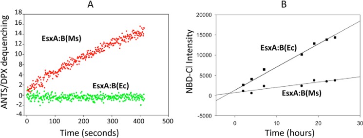
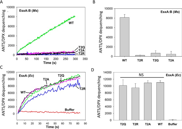
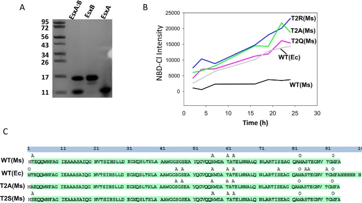
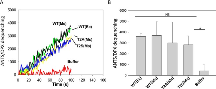
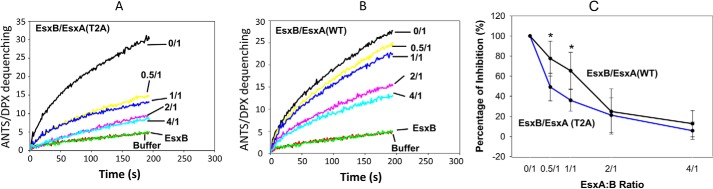
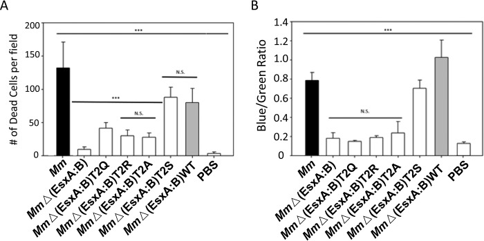
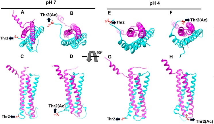
The Mycobacterium tuberculosis virulence factor EsxA and its chaperone EsxB are secreted as a heterodimer (EsxA:B) and are crucial for mycobacterial escape from phagosomes and cytosolic translocation. Current findings support the idea that for EsxA to interact with host membranes, EsxA must dissociate from EsxB at low pH. However, the molecular mechanism by which the EsxA:B heterodimer separates is not clear. In the present study, using liposome-leakage and cytotoxicity assays, LC-MS/MS-based proteomics, and CCF-4 FRET analysis, we obtained evidence that the Nα-acetylation of the Thr-2 residue on EsxA, a post-translational modification that is present in mycobacteria but absent in Escherichia coli, is required for the EsxA:B separation. Substitutions at Thr-2 that precluded Nα-acetylation inhibited the heterodimer separation and hence prevented EsxA from interacting with the host membrane, resulting in attenuated mycobacterial cytosolic translocation and virulence. Molecular dynamics simulations revealed that at low pH, the Nα-acetylated Thr-2 makes direct and frequent "bind-and-release" contacts with EsxB, which generates a force that pulls EsxB away from EsxA. In summary, our findings provide evidence that the Nα-acetylation at Thr-2 of EsxA facilitates dissociation of the EsxA:B heterodimer required for EsxA membrane permeabilization and mycobacterial cytosolic translocation and virulence.

Keywords: ESAT-6; EsxA; Mycobacterium tuberculosis; Nα-acetylation; bacterial pathogenesis; chaperone; membrane; post-translational modification (PTM); protein-protein interaction; virulence factor.


Conflict of interest statement

The authors declare that they have no conflicts of interest with the contents of this article

Figures

Figure 1.
Figure 1.
The EsxA:B heterodimer produced in Ms, but not in E. coli, permeabilized the liposomes at low pH, implicating the role of Nα-acetylation in heterodimer dissociation. A, the heterodimers EsxA:B purified from M. smegmatis (Ms) or E. coli (Ec) were tested in three independent experiments with ANTS/DPX dequenching assay at pH 4.0. B, equal amounts of the heterodimer EsxA:B(Ms) or EsxA:B(Ec) were incubated with NBD-Cl at room temperature. NBD-Cl is a fluorescent dye that only reacts with a free N-terminal site without Nα-acetylation was recorded at 535 nm (excitation at 480 nm) and plotted as a function of time.
Figure 2.
Figure 2.
The mutations at Thr-2 of EsxA diminished the membrane-permeabilizing activity of EsxA:B heterodimer. A, the EsxA:B heterodimer proteins (WT and the mutants carrying mutations T2A, T2Q, and T2R) were purified from Ms. The membrane-permeabilizing activity of the purified heterodimer proteins was tested by ANTS/DPX fluorescence dequenching assay. The representative ANTS/DPX fluorescent dequenching curves were shown. B, the average end-point fluorescence intensities from at least three independent experiments were calculated. The results represent the averages of three replicates, and error bars represent S.D. C, the EsxA proteins (WT and T2A, T2Q, and T2R mutants) were purified from Ec. The membrane-permeabilizing activity of the Ec-EsxA proteins purified from Ec was tested by ANTS/PDX assay. Representative curves are shown. D, the average end-point fluorescence intensities from at least three independent experiments were calculated, with error bars denoting S.D. NS, not significant.
Figure 3.
Figure 3.
Detection of Nα-acetylation of EsxA by NBD-Cl and MS. A, the EsxA:B heterodimer purified from Ms was separated by 6 m guanidine, after which EsxA and EsxB were purified separately via nickel affinity as described under “Experimental procedures.” B, the indicated EsxA proteins, purified from Ms, were incubated with NBD-Cl. At the indicated times, the fluorescence intensity of NBD-Cl was measured. C, the indicated EsxA proteins, purified from Ms or Ec as indicated, were analyzed via LC-MS/MS to identify the post-translational modifications. The residues with acetylation are labeled as A, and the residues with oxidation are labeled as O.
Figure 4.
Figure 4.
The Nα-acetylation of EsxA did not affect the membrane-permeabilizing activity of EsxA. A, the indicated Ms-EsxA proteins isolated from the Ms-heterodimer were tested for membrane-permeabilizing activity using ANTS/DPX assay. The Ec-EsxA (WT) protein, purified from Ec, was used as a control. The representative curves from at least three independent experiments are shown. B, the average end-point fluorescence intensity from at least three independent experiments was calculated and is shown (p < 0.05).
Figure 5.
Figure 5.
EsxB preferentially inhibited nonacetylated EsxA(T2A) over its Nα-acetylated counterpart. The Ms-produced EsxA(WT) and EsxA(T2A) proteins were incubated with various concentrations of EsxB as the indicated molar ratios. The mixtures were tested in triplicate by ANTS/DPX assay for membrane-permeabilizing activity. The representative curves are shown in A and B, respectively. The relative inhibition from at least three independent experiments for a total n = 9 was summarized in C. For EsxA:B 0.5:1.0 ratio, p = 0.0015 and for EsxA:B 1:1 ratio p = 0.0012. Error bars represent S.D.
Figure 6.
Figure 6.
The non-Nα-acetylate EsxA diminished mycobacterial virulence and cytosolic translocation in macrophages. A, RAW263.4 cells were infected with the indicated Mm strains at MOI of 10. The cytotoxicity was measured by using the live/dead assay. Dead cells were counted in random fields (***, n = 22, p < 0.0001). B, mycobacterial cytosolic translocation was monitored by using CCF4-AM as a FRET reporter. The blue/green ratio was measured by comparing emissions at 450 and 530 nm with excitation at 409 nm. The data were calculated from at least three independent experiments (***, n = 3, p < 0.0001).
Figure 7.
Figure 7.
Molecular dynamic simulation detects the acetylated Thr-2(Ac) interacts with EsxB in a bind-and-release mode. The structures of Mtb EsxA:B heterodimers with/out Nα-acetylation were analyzed by molecular dynamic simulation. The figures were generated from snapshots of 20-ns MD simulations at pH 7 and pH 4. EsxA, EsxB, and the Thr-2 residue are shown in cyan, pink, and red, respectively. The structures of EsxA:B heterodimer with nonacetylated Thr-2 at pH 7 and pH 4 are shown in A (pH 7, top view), C (pH 7, side view), E (pH 4, top view), and G (pH 4, side view), respectively. The structures of EsxA:B heterodimer with acetylated Thr-2(Ac) are shown in B (pH 7, top view), D (pH 7, side view), F (pH 4, top view), and H (pH 4, side view), respectively.

References

    1. Zumla A., Raviglione M., Hafner R., and von Reyn C. F. (2013) Current concepts. N. Engl. J. Med. 368, 745–755 10.1056/NEJMra1200894 - DOI - PubMed
    1. Global Tuberculosis Report 2018 (2018) World Health Organization, Geneva, Switzerland
    1. Orme I. (2004) Adaptive immunity to mycobacteria. Curr. Opin. Microbiol. 7, 58–61 10.1016/j.mib.2003.11.002 - DOI - PubMed
    1. Kang P. B., Azad A. K., Torrelles J. B., Kaufman T. M., Beharka A., Tibesar E., DesJardin L. E., and Schlesinger L. S. (2005) The human macrophage mannose receptor directs Mycobacterium tuberculosis lipoarabinomannan-mediated phagosome biogenesis. J. Exp. Med. 202, 987–999 10.1084/jem.20051239 - DOI - PMC - PubMed
    1. Pizarro-Cerdá J., and Cossart P. (2006) Bacterial adhesion and entry into host cells. Cell 124, 715–727 10.1016/j.cell.2006.02.012 - DOI - PubMed

Publication types

MeSH terms

Substances

Associated data