. 2020 Mar 31;117(13):7296-7304.
doi: 10.1073/pnas.1920860117. Epub 2020 Mar 13.

Hox genes maintain critical roles in the adult skeleton

Affiliations

Hox genes maintain critical roles in the adult skeleton

Jane Y Song et al. Proc Natl Acad Sci U S A. .

Abstract

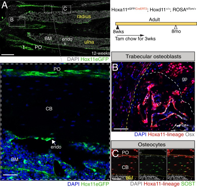
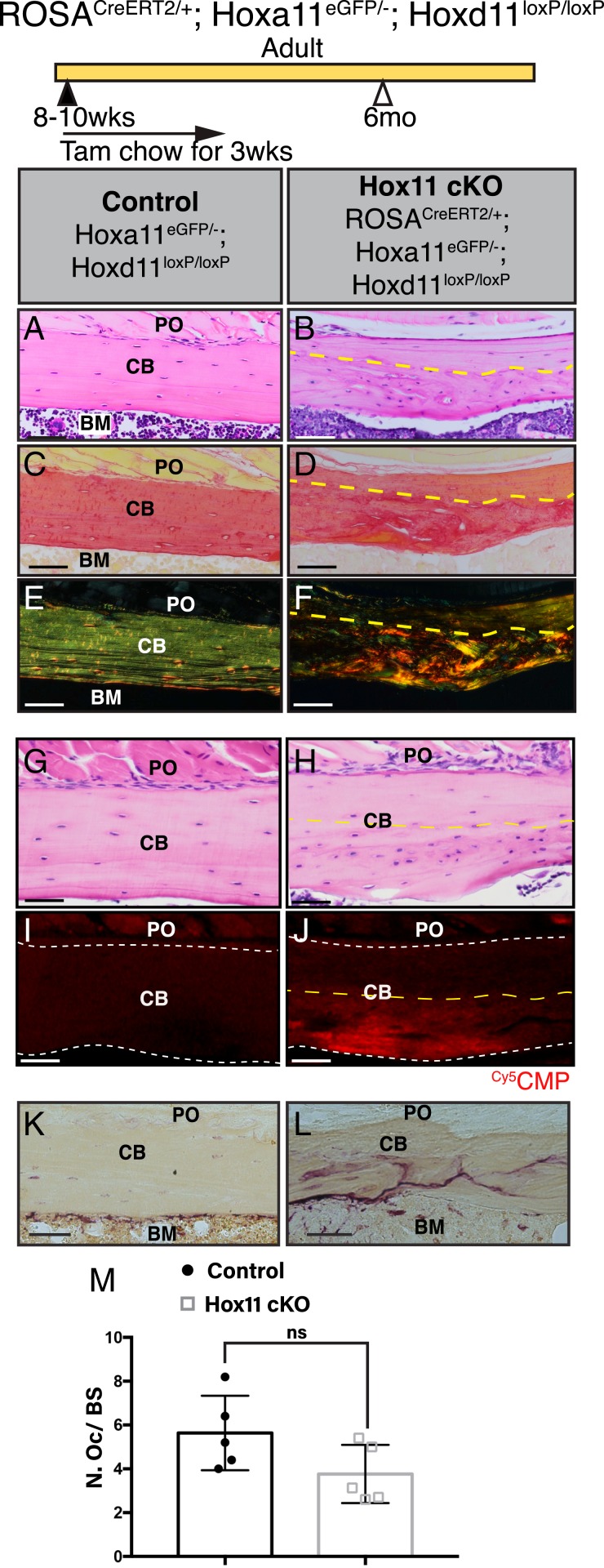
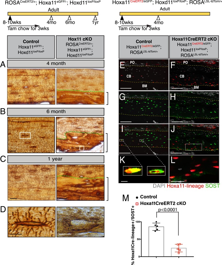
Hox genes are indispensable for the proper patterning of the skeletal morphology of the axial and appendicular skeleton during embryonic development. Recently, it has been demonstrated that Hox expression continues from embryonic stages through postnatal and adult stages exclusively in a skeletal stem cell population. However, whether Hox genes continue to function after development has not been rigorously investigated. We generated a Hoxd11 conditional allele and induced genetic deletion at adult stages to show that Hox11 genes play critical roles in skeletal homeostasis of the forelimb zeugopod (radius and ulna). Conditional loss of Hox11 function at adult stages leads to replacement of normal lamellar bone with an abnormal woven bone-like matrix of highly disorganized collagen fibers. Examining the lineage from the Hox-expressing mutant cells demonstrates no loss of stem cell population. Differentiation in the osteoblast lineage initiates with Runx2 expression, which is observed similarly in mutants and controls. With loss of Hox11 function, however, osteoblasts fail to mature, with no progression to osteopontin or osteocalcin expression. Osteocyte-like cells become embedded within the abnormal bony matrix, but they completely lack dendrites, as well as the characteristic lacuno-canalicular network, and do not express SOST. Together, our studies show that Hox11 genes continuously function in the adult skeleton in a region-specific manner by regulating differentiation of Hox-expressing skeletal stem cells into the osteolineage.

Keywords: Hox genes; MSCs; bone matrix; osteolineage differentiation; skeletal homeostasis.


Conflict of interest statement

The authors declare no competing interest.

Figures

Fig. 1.
Fig. 1.
Adult Hox11-expressing skeletal stem cells continuously give rise to osteoblasts and osteocytes. (A) Hoxa11-eGFP real-time reporter allele demonstrates continuous expression of Hoxa11 (green) at 12 wk of age. The panel was created by stitching 17 individual 10× images. t, tendon. DAPI, gray. (Scale bar: 200 μm.) Shown is a higher magnification image of the boxed area in A showing localization of Hoxa11 expression (green) in the periosteum, endosteum, and bone marrow compartment. BM, bone marrow; CB, cortical bone; endo, endosteum; PO, periosteum. DAPI, blue. (Scale bar: 75 μm.) Animals of the indicated genotype were fed on tamoxifen (Tam) chow at 8 to 10 wk of age for a duration of 3 wk to induce deletion and collected at 8 mo of age. Boxes marked “B” and “C” in A represent the approximate locations of magnified images shown in B and C. (B) Hoxa11-lineage marked cells (red) are found in the trabecular bone coexpressing osterix (Osx) (white). The yellow dashed line outlines the cortical bone, and the white dashed line demarcates the growth plate border. gp, growth plate. DAPI, blue. (Scale bar: 75 μm.) (C) Hoxa11-lineage marked cells (red) are also found as osteocytes embedded within the cortical bone coexpressing SOST (green). Hoxa11-lineage marked cells and DAPI (gray) in the far-left, SOST (green) and DAPI (gray) in the middle, and the merged image is shown in the far-right. (Scale bar: 60 μm.) All images: BM, bone marrow; CB, cortical bone; endo, endosteum; PO, periosteum.
Fig. 2.
Fig. 2.
Conditional deletion of Hox11 function recapitulates the germline null mutation. (A) Schematic illustrating the Hoxd11 locus. Yellow arrowheads illustrate the inserted loxP sites. Two guide RNAs with the indicated sequences (underlined) along with their corresponding PAM (highlighted blue) were used to flank exon 2 of Hoxd11 in order to insert loxP sites. Homology sequences used in the donor sequences are highlighted with thick dark blue line (5′ loxP) and thick light blue line (3′ loxP). Red arrows marked with P1, P2, and P3 mark the location of the PCR primers used to confirm recombination. Corresponding PCR product sizes are indicated as well. The PCR elongation time was adjusted so that a 300-bp PCR product would appear only if recombination had occurred between the loxP sites. Pregnant dams were fed on tamoxifen chow for 1 wk to induce recombination, and the resulting embryos were collected at E17.5. (B) PCR analysis using the PCR primers produces a robust 600-bp control band only present in the controls and a 300-bp recombined band only present in the conditional mutants. cKO denotes Hox11 conditional mutants. Skeletal preparations of limbs from (C) WT, (D) littermate control for Hox11 conditional mutant, (E) Hox11ROSACreERT2 conditional mutant, and (F) Hox11 germline-null mutant. The red box highlights the zeugopod skeleton.
Fig. 3.
Fig. 3.
Deletion of Hox11 function at adult stages results in the regional disruption in cortical bone homeostasis. Hox11 conditional mutants with the ROSA-CreERT2 (genotype indicated) allele along with control animals were fed on tamoxifen chow beginning at 8 to 10 wk of age for a total of 3 wk to delete Hox11 function. Animals were evaluated at 4 mo of age, 6 mo of age, and 1 y of age. (A) Tail samples from all animals collected were analyzed via PCR to assess recombination. A robust 300-bp band in the conditional mutants demonstrates strong recombination. (B) qRT-PCR of Hoxa11eGFP-expressing zeugopod skeletal cells from the conditional mutants show robust deletion of Hoxd11 in the conditional mutants. Data are presented relative to mouse GAPDH using the ΔΔCt method. ND, none detected. Error is represented as mean ± SEM. (CH) H&E stains of paraffin bone sections (ulna) of control and Hox11ROSACreERT2conditional mutant animals. The dashed line demarcates the border between lamellar (above) and abnormal (below) bone. Green brackets demarcate the abnormal matrix. Boxed areas in D and G represent high-magnification images of an individual matrix-embedded cell highlighting the morphological differences between control and Hox11 conditional mutant osteocyte-like cells. (I) Quantification of cells embedded within the abnormal matrix at 4 mo, 6 mo, and 1 y of age shows a significant increase in cell number in Hox11ROSACreERT2 conditional mutant bone. Error is represented as mean ± SEM. Statistics by Student’s t test. (J) Percentage of osteocytes or osteocyte-like cells in Hox11 conditional mutant bones that exhibit an ellipsoid shape within lamellar (above dotted line in FH) and abnormal (below dotted line, green bracket in FH) bony matrix. Error is represented as mean ± SEM. Statistics by Student’s t test. (K and L) H&E stains of bones from the humerus of control (K) and Hox11 conditional mutant (L) show no differences in morphology at 1 y of age. All images: BM, bone marrow; CB, cortical bone; PO, periosteum. (Scale bar in all images: 100 μm.)
Fig. 4.
Fig. 4.
Woven bone-like region in the Hox11 conditional mutant bones has a disorganized collagen matrix. Hox11 conditional mutants with the ROSA-CreERT2 (genotype indicated) allele along with control animals were fed on tamoxifen chow for 3 wk beginning at 8 to 10 wk of age to induce deletion, and animals were examined at 6 mo of age. (A and B) H&E stains of paraffin-processed bone sections of control and Hox11ROSACreERT2 conditional mutant animals. (C and D) Brightfield images of picrosirius red stain of consecutive bone sections from A and B. (E and F) Polarized light images of picrosirius red stain of bone sections from C and D. (G and H) H&E stains of paraffin-processed bone sections of control and Hox11ROSACreERT2 conditional mutant animals. (I and J) Consecutive bone sections from G and H stained with Cy5CMP (red). White dashed line marks border of cortical bone. (K and L) Control (K) and Hox11ROSACreERT2 conditional mutant (L) bone sections stained with TRAP. Note the distinct distribution of TRAP stain in conditional mutant. (M) Quantification of osteoclast number (N. Oc) on bone surface (BS) using the Bioquant Osteo software. Statistics by Student’s t test. Error is represented as mean ± SEM. All images are from the ulna. BM, bone marrow; CB, cortical bone; PO, periosteum. The yellow dashed line demarcates border between lamellar (above) and abnormal (below) bone. ns, not significant. (Scale bar in all images: 100 μm.)
Fig. 5.
Fig. 5.
Osteoblast differentiation is perturbed in Hox11 conditional mutant bone. Hox11 conditional mutants with the Hoxa11-CreERT2 allele (genotype indicated) along with control animals were fed on tamoxifen chow for 3 wk starting at 8 to 10 wk of age and collected at 4 mo of age. (A) Brightfield image of a bone section from a control animal overlaid with Hox11-lineage marked cells (red) shows contribution to osteocytes. (B) Brightfield image of bone section from a Hox11Hoxa11CreERT2 conditional mutant overlaid with Hox11-lineage–positive cells (red) shows contribution to abnormal bone matrix. (C and D) Control (C) and Hox11Hoxa11CreERT2 conditional mutant (D) bone stained with RUNX2 (magenta). DAPI, gray. White arrows in C and D mark the endosteal bone surfaces. (E and F) Control (E) and Hox11Hoxa11CreERT2 conditional mutant (F) bone stained with osteopontin (Opn, green). (G and H) Control (G) and Hox11Hoxa11CreERT2 conditional mutant (H) bone sections stained with osteocalcin (Ocn, green). (IJ″) Hoxa11eGFP (green) and DAPI (blue) in I and J, Hoxa11-lineage marked cells (red) and DAPI (blue) in I′ and J′, and merged images in I″ and J″. Control (II″) and Hox11Hoxa11CreERT2 conditional mutant (JJ″) bone sections show Hoxa11-lineage marked (red), non-Hoxa11eGFP endosteal surface osteoblasts (yellow arrowhead). Notice stark difference in morphology. All images are from the ulna. DAPI, blue (unless noted otherwise); Hox11-lineage marked cells, red. CB, cortical bone; endo, endosteum. The white dashed line demarcates the endosteal surface. (Scale bars in all images: 50 μm.)
Fig. 6.
Fig. 6.
Hox11 conditional mutant osteocytes fail to form dendrites or express SOST. Hox11 conditional mutants with the ROSA-CreERT2 (genotype indicated) allele along with control animals were fed on tamoxifen chow beginning at 8 to 10 wk of age for 3 wk to delete Hox11 function, and animals were evaluated at 4 mo of age, 6 mo of age, and 1 y of age for CE. (AC) Control (Left) and Hox11ROSACreERT2 conditional mutants (Right) were treated with silver nitrate. Green dashed lines demarcate the lamellar (above) and abnormal (below) bone. Brackets outline abnormal bone matrix. (D) High magnification of osteocytes from the white-boxed area in B. Hox11 conditional mutants with the Hoxa11-CreERT2 allele (genotype indicated) along with control animals were fed on tamoxifen chow for 3 wk starting at 8 to 10 wk of age and collected at 4 mo of age for EM. (E and F) Control (E) and Hox11Hoxa11CreERT2 conditional mutant (F) bone sections showing Hox11-lineage marked cells (red) that contributed to osteocytes. (G and H) Control (G) and Hox11Hoxa11CreERT2 conditional mutant (H) bone sections stained with SOST (green). (I and J) Merged images of E and G in I or F and H in J showing overlap or the lack thereof of Hox11-lineage marked cells (red) and SOST (green). The yellow dashed line in EJ outline the cortical bone. (K and L) Close-up of osteocytes in white-boxed region in I and J. (M) Quantification of Hox11-lineage marked cells (red) that also express SOST (green). Error is represented as mean ± SEM. Statistics by Student’s t test. All images are from the ulna. DAPI, gray. BM, bone marrow; CB, cortical bone; PO, periosteum. (Scale bar: 25 μm [AC], 10 μm [D], 100 μm [EJ], 10 μm [K and L].)

References

    1. Mallo M., Wellik D. M., Deschamps J., Hox genes and regional patterning of the vertebrate body plan. Dev. Biol. 344, 7–15 (2010). - PMC - PubMed
    1. Fromental-Ramain C., et al. , Specific and redundant functions of the paralogous Hoxa-9 and Hoxd-9 genes in forelimb and axial skeleton patterning. Development 122, 461–472 (1996). - PubMed
    1. Fromental-Ramain C., et al. , Hoxa-13 and Hoxd-13 play a crucial role in the patterning of the limb autopod. Development 122, 2997–3011 (1996). - PubMed
    1. Wellik D. M., Capecchi M. R., Hox10 and Hox11 genes are required to globally pattern the mammalian skeleton. Science 301, 363–367 (2003). - PubMed
    1. Davis A. P., Witte D. P., Hsieh-Li H. M., Potter S. S., Capecchi M. R., Absence of radius and ulna in mice lacking hoxa-11 and hoxd-11. Nature 375, 791–795 (1995). - PubMed

Publication types

LinkOut - more resources