. 2020 Mar 13;11(1):1397.
doi: 10.1038/s41467-020-15230-y.

Exploration of sensory and spinal neurons expressing gastrin-releasing peptide in itch and pain related behaviors

Affiliations

Exploration of sensory and spinal neurons expressing gastrin-releasing peptide in itch and pain related behaviors

Devin M Barry et al. Nat Commun. .

Abstract

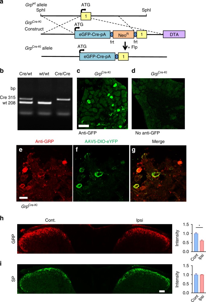
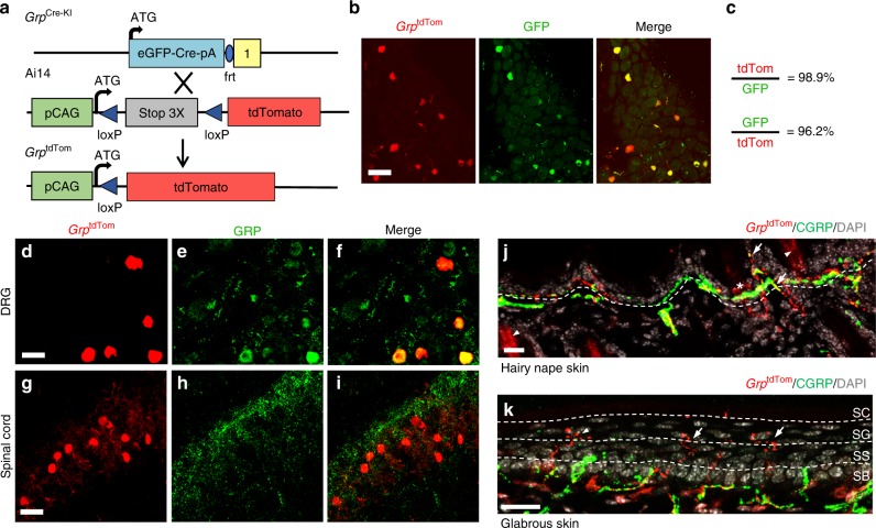
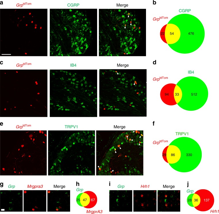
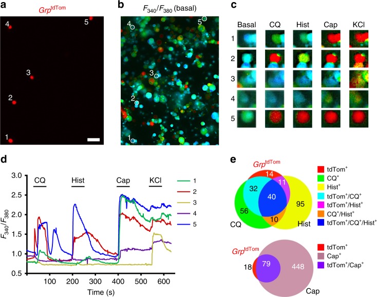
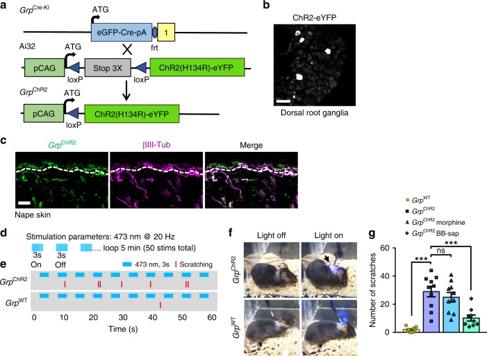
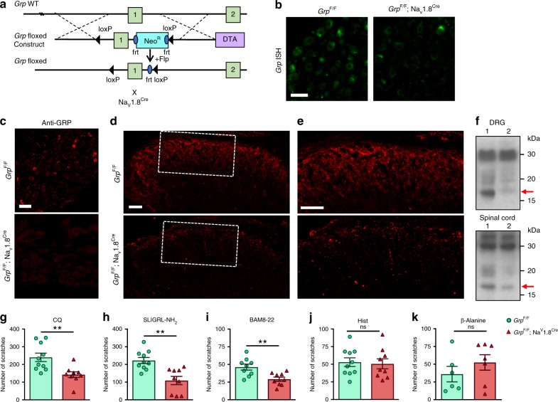
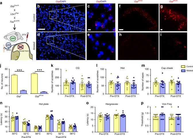
Gastrin-releasing peptide (GRP) functions as a neurotransmitter for non-histaminergic itch, but its site of action (sensory neurons vs spinal cord) remains controversial. To determine the role of GRP in sensory neurons, we generated a floxed Grp mouse line. We found that conditional knockout of Grp in sensory neurons results in attenuated non-histaminergic itch, without impairing histamine-induced itch. Using a Grp-Cre knock-in mouse line, we show that the upper epidermis of the skin is exclusively innervated by GRP fibers, whose activation via optogeneics and chemogenetics in the skin evokes itch- but not pain-related scratching or wiping behaviors. In contrast, intersectional genetic ablation of spinal Grp neurons does not affect itch nor pain transmission, demonstrating that spinal Grp neurons are dispensable for itch transmission. These data indicate that GRP is a neuropeptide in sensory neurons for non-histaminergic itch, and GRP sensory neurons are dedicated to itch transmission.


Conflict of interest statement

The authors declare no competing interests.

Figures

Fig. 1
Fig. 1. Validated expression of Grp and Cre in GrpCre-KI sensory neurons.
a Schematic of targeting strategy to knock in eGFP-Cre cassette in Grp allele to generate GrpCre-KI mice. b Gel electrophoresis of genotyping PCR from Grp Cre/wt, wt/wt, and Cre/Cre samples. c eGFP IHC from DRG section of an adult GrpCre-KI mouse. d Image of eGFP epifluorescent signal (no GFP antibody) in GrpCre-KI DRG. eg GRP IHC (e) and eYFP fluorescent image (f) from DRG section of an adult GrpCre-KI mouse that received AAV5- EF1α-DIO-eYFP injection into the DRG for 3 weeks. Scale bar, 20 μm. h and i IHC of GRP (h) and SP (i) in cervical spinal sections from WT mice following i.d. injections of CQ (200 μg). Scale bar, 100 μm. Data are presented as mean ± s.e.m., n = 3 mice and 9 sections. Source data are provided as a Source Data file.
Fig. 2
Fig. 2. GrptdTom recapitulates Grp expression in DRG and the spinal cord.
a Schematic of GrpCre-KI mating with Ai14 reporter line to produce GrptdTom mice. b IHC images of eGFP and tdTomato epifluorescence in GrptdTom DRG. Scale bar, 50 μm. c Percentage of eGFP+ neurons co-expressing tdTomato or of tdTomato+ neurons co-expressing eGFP. n = 3 mice and 10 sections. df IHC images of tdTomato epifluorescence (d) and GRP (e) in GrptdTom DRG. Scale bar, 20 μm. gi IHC of tdTomato epifluorescence (g) and GRP (h) in GrptdTom spinal cord. Scale bars, 50 µm. j IHC image of tdTomato, CGRP and DAPI in GrptdTom nape skin. Arrows indicate apparent GrptdTom fibers co-expressing CGRP. Arrowheads indicate autofluorescent hair shafts. Dashed line indicates epidermal/dermal boundary. k IHC images of tdTomato, CGRP, and DAPI in glabrous paw skin with stratum basilis (SB), stratum spinosum (SS), stratum granulosum (SG), and stratum corneum (SC) epidermal layers marked by dashed lines. Arrows indicate GrptdTom fibers with ‘S’ or ‘Z’ pattern nerve endings. Arrowhead indicates GrptdTom fibers with apparent bush endings. Scale bars, 20 μm.
Fig. 3
Fig. 3. Grp expression is enriched in a subset of sensory neurons with both peptidergic and non-peptidergic markers.
a, c, e IHC image of tdTomato with CGRP (a), IB4 (c), and TRPV1 (e) in GrptdTom DRG. Arrowheads indicate double stained neurons. Scale bar, 50 μm. b, d, f Venn Diagram of DRG neurons with tdTomato/CGRP expression, tdTomato/IB4-binding, and tdTomato/TRPV1 expression from GrptdTom mice. g ISH of Grp and Mrgpra3 in WT DRG. Scale bar, 20 μm. h Diagram of DRG neurons with Grp and Mrgpra3 expression. i ISH of Grp and Hrh1 in WT DRG. j Diagram of DRG neurons with Grp and Hrh1 expression. n = 3 mice and 9 sections.
Fig. 4
Fig. 4. Pruritogens induce Ca2+ responses in Grp+ sensory neurons.
a and b Image of tdTomato neurons (a) and F340 / F380 signal (b) from GrptdTom DRG cultures loaded with fura 2-AM. Scale bar, 50 μm. c Snapshots of GrptdTom neuron intracellular Ca2+ levels at basal and 20 s after CQ (1 mM), Hist (100 μM), Cap (100 nM) or KCl (30 mM) applications. d F340/F380 traces from individual GrptdTom neurons with application of CQ, Hist, Cap and KCl. e Venn diagram of GrptdTom DRG neurons responsive to CQ, Hist or Cap and/or expressing tdTomato. n = 4 mice and 1424 neurons for ae. Source data are provided as a Source Data file.
Fig. 5
Fig. 5. Opto-activation of Grp+ sensory neuron skin fibers evokes itch behavior.
a Schematic of GrpCre-KI mating with Ai32 ChR2-eYFP line to produce GrpChR2 mice. b IHC Image of eYFP expression in GrpChR2 DRG. Scale bar, 50 μm. c IHC images of eYFP and βIII-Tubulin in GrpChR2 nape skin. Dashed lines indicate epidermal/dermal boundary. Scale bar, 100 μm. d Optical parameters of skin fiber stimulation GrpChR2 and GrpWT mice. e Raster plot of scratching behavior induced by light stimulation of skin in GrpChR2 and GrpWT mice. f Snapshots of GrpChR2 and GrpWT mice with light off or on. Arrow indicates hind paw scratching the nape when light is on. g Total number of scratches during 5-min light stimulation experiment in GrpWT, GrpChR2, GrpChR2 morphine-treated and GrpChR2 BB-sap-treated mice. Data are presented as mean ± s.e.m., n = 8 mice for GrpWT, 9 mice for GrpChR2 BB-sap, and 10 mice for GrpChR2 and GrpChR2 morphine, one-way ANOVA with Tukey post hoc, ***p < 0.001, ns not significant. Source data are provided as a Source Data file.
Fig. 6
Fig. 6. Attenuated itch behaviors in mice with conditional deletion of Grp in sensory neurons.
a Schematic of targeting strategy for inserting loxP sites into Grp allele to generate Grp floxed (GrpF/F) mice and subsequent crossing with Nav1.8Cre mice. b ISH images of Grp expression in DRG sections from GrpF/F and GrpF/F; Nav1.8Cre mice. Scale bar, 50 μm. c IHC images of GRP antibody in DRG sections from GrpF/F and GrpF/F; Nav1.8Cre mice. Scale bar, 50 μm. d IHC images of GRP antibody in spinal cord sections from GrpF/F and GrpF/F; Nav1.8Cre mice. Scale bar, 100 μm. e High power images of boxed area in d. Scale bar, 100 μm. f Western blot of GRP in DRG (upper row) and the spinal cord (lower row) from GrpF/F (lane 1) and GrpF/F; Nav1.8Cre mice (lane 2). Arrow, predicted size of pro-GRP at 15.6 kDa. gk Mean number of scratches induced by i.d. nape injection of (g) CQ (200 μg), (h) SLIGRL-NH2 (100 μg), (i) BAM8-22 (100 μg), (j) Hist (200 μg), and (k) β-alanine (100 µg) in GrpF/F and GrpF/F; Nav1.8Cre littermates. Data are presented as mean ± s.e.m., n = 9 mice for GrpF/F and 10 mice for GrpF/F;Nav1.8Cre in gj. n = 6 mice for GrpF/F and 7 mice for GrpF/F;Nav1.8Cre in k. **p < 0.01, unpaired two tails t test in gk. Source data are provided as a Source Data file.
Fig. 7
Fig. 7. Intersectional ablation of Grp spinal neurons does not affect itch or pain behavior responses.
a Schematic of mating strategy of GrpCre-KI with Lbx1Flpo and Tauds-DTR lines to generate DTR-expressing Grp spinal neurons and subsequent DTX injection to ablate Grp spinal neurons. be ISH images of Grp expression in cervical spinal cord sections from (b, c) control and (d, e) ablated littermates. Scale bars, 100 μm in b and 20 μm in c. fi Epifluorescent images of tdTomato in cervical spinal cord sections from (f, g) GrptdTom control and (h, i) GrptdTom ablated mice. Scale bars, 100 μm in f and 20 μm in g. j Mean number of Grp + neurons and GrptdTom neurons in control and ablated sections. n = 3 mice and 10 sections, unpaired t test, ***p < 0.001. k, l Mean number of scratches induced by i.d. nape injection of (k) CQ (100 μg) and (l) Hist (200 μg) in control and ablated mice pre- and post-DTX injection. m Mean number of wipes induced by i.d. cheek injection of Cap (20 μg) in control and ablated mice pre- and post-DTX injection. n Mean response latencies for hot plate assay at 50 °C, 52 °C, and 56 °C in control and ablated mice pre- and post-DTX injection. o Mean response latencies for Hargreaves hind-paw assay in control and ablated mice pre- and post-DTX injection. p Withdrawal thresholds (in grams of force, g) for Von Frey filament assay in control and ablated mice pre- and post-DTX injection. n = 10 mice, two-way RM ANOVA with Tukey post-hoc in kp. Data are presented as mean ± s.e.m. Source data are provided as a Source Data file.

Similar articles

Cited by

References

    1. Barry DM, Munanairi A, Chen ZF. Spinal mechanisms of itch transmission. Neurosci. Bull. 2017;34:146–164. - PMC - PubMed
    1. Bautista DM, Wilson SR, Hoon MA. Why we scratch an itch: the molecules, cells and circuits of itch. Nat. Neurosci. 2014;17:175–182. doi: 10.1038/nn.3619. - DOI - PMC - PubMed
    1. Jeffry J, Kim S, Chen ZF. Itch signaling in the nervous system. Physiology. 2011;26:286–292. doi: 10.1152/physiol.00007.2011. - DOI - PMC - PubMed
    1. Braz J, Solorzano C, Wang X, Basbaum AI. Transmitting pain and itch messages: a contemporary view of the spinal cord circuits that generate gate control. Neuron. 2014;82:522–536. doi: 10.1016/j.neuron.2014.01.018. - DOI - PMC - PubMed
    1. Sun YG, Chen ZF. A gastrin-releasing peptide receptor mediates the itch sensation in the spinal cord. Nature. 2007;448:700–703. doi: 10.1038/nature06029. - DOI - PubMed

Publication types

MeSH terms