. 2020 Mar 13;11(1):1334.
doi: 10.1038/s41467-020-14957-y.

Extracellular nanovesicles for packaging of CRISPR-Cas9 protein and sgRNA to induce therapeutic exon skipping

Affiliations

Extracellular nanovesicles for packaging of CRISPR-Cas9 protein and sgRNA to induce therapeutic exon skipping

Peter Gee et al. Nat Commun. .

Abstract

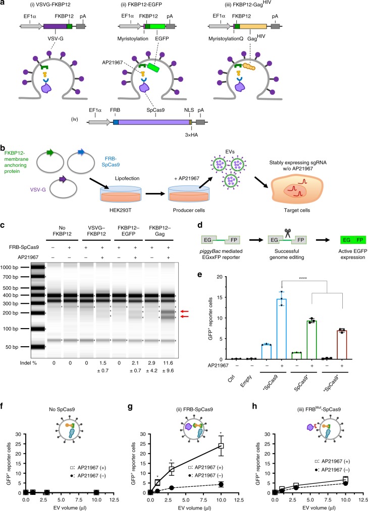
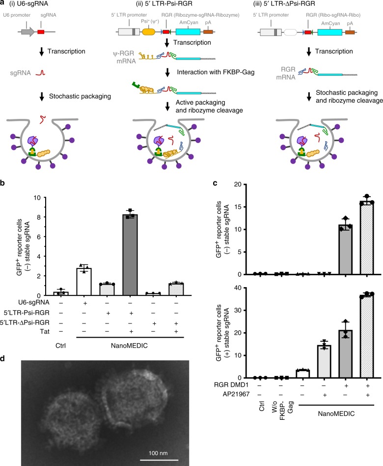
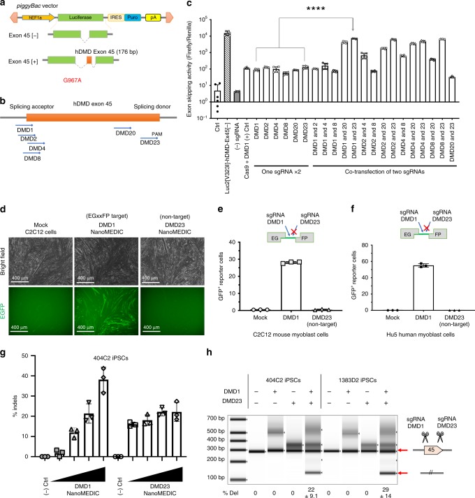
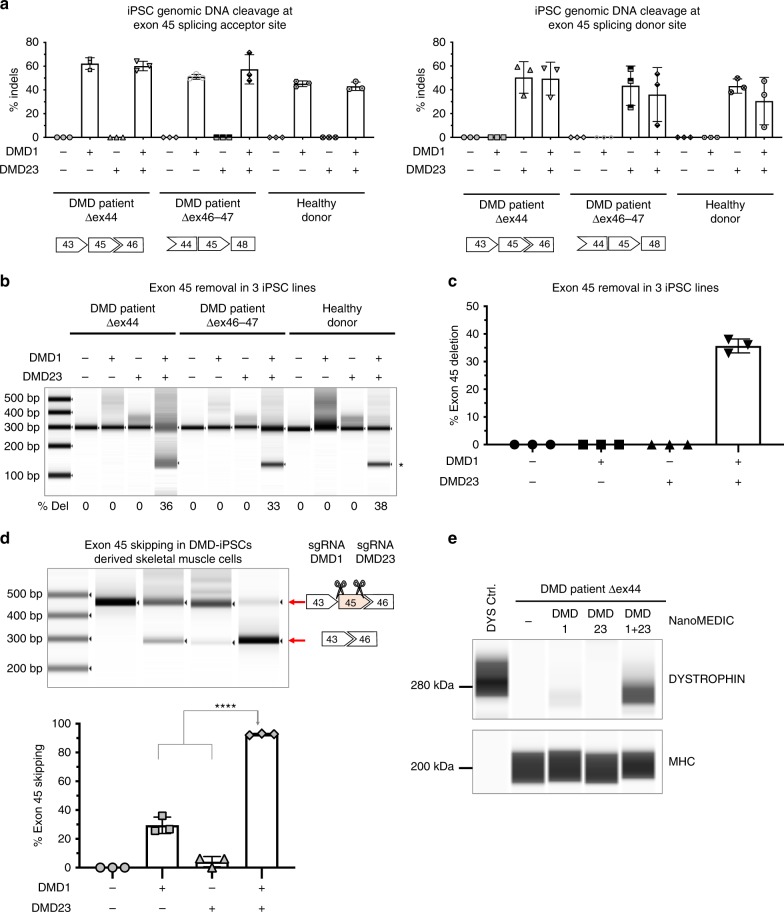
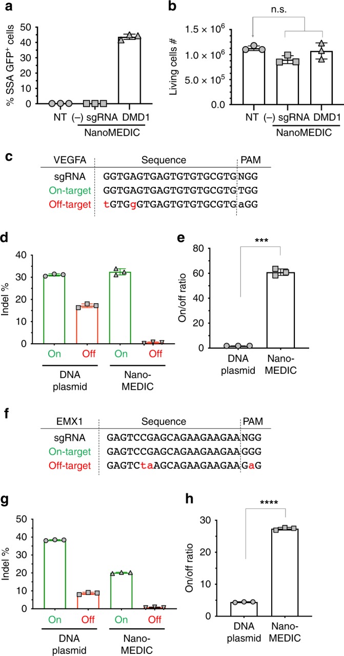
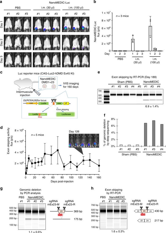
Prolonged expression of the CRISPR-Cas9 nuclease and gRNA from viral vectors may cause off-target mutagenesis and immunogenicity. Thus, a transient delivery system is needed for therapeutic genome editing applications. Here, we develop an extracellular nanovesicle-based ribonucleoprotein delivery system named NanoMEDIC by utilizing two distinct homing mechanisms. Chemical induced dimerization recruits Cas9 protein into extracellular nanovesicles, and then a viral RNA packaging signal and two self-cleaving riboswitches tether and release sgRNA into nanovesicles. We demonstrate efficient genome editing in various hard-to-transfect cell types, including human induced pluripotent stem (iPS) cells, neurons, and myoblasts. NanoMEDIC also achieves over 90% exon skipping efficiencies in skeletal muscle cells derived from Duchenne muscular dystrophy (DMD) patient iPS cells. Finally, single intramuscular injection of NanoMEDIC induces permanent genomic exon skipping in a luciferase reporter mouse and in mdx mice, indicating its utility for in vivo genome editing therapy of DMD and beyond.


Conflict of interest statement

P.G. and A.H. are inventors on a filed patent (PCT/JP2019/027708) based on the delivery system, virus-like particle, production, and use for genome editing described here. Y.M., H.H., and N.I. are employees of Takeda Pharmaceutical Company. A.H. is a principle investigator (without salary) of the T-CiRA program funded by Takeda Pharmaceutical Company. All other authors declare no competing interests.

Figures

Fig. 1
Fig. 1. Selective packaging of SpCas9 protein into NanoMEDIC by chemical-induced dimerization.
a Schematics of membrane-anchoring constructs fused with FKBP12 and FRB-SpCas9. b Schematic of NanoMEDIC production from producer cells and delivery into recipient cells. EGFP is neon green. The myristoylation domain is indicated in pink. The FKBP12 heterodimerization domain is dark green and the FRB heterodimerization domain is blue. GagHIV is tan. SpCas9 protein is light purple. The dimerization ligand, AP21967, is yellow. VSV-G envelope is dark purple and on the surface of the cell. c T7E1 analysis of HEK293T cells stably expressing sgRNA targeting DMD1. These cells were inoculated with NanoMEDIC containing FRB-SpCas9 and no FKBP12 interaction partner, VSVG-FKBP12, FKBP12-EGFP-A, or FKBP12-GagHIV, produced in the presence or absence of AP21967. Red arrowheads show cleaved products by T7E1enzyme. Data are mean ± S.D. from technical triplicates. d Schematic showing NanoMEDIC inoculation onto HEK293T EGxxFP reporter cells stably expressing DMD1-sgRNA and the resulting GFP expression upon cleavage by delivered SpCas9 protein. e HEK293T EGxxFP reporter cells stably expressing sgRNA were inoculated with NanoMEDIC containing FKBP12-GagHIV and FRB-SpCas9 (*SpCas9), SpCas9-FRB (SpCas9*), or FRB-SpCas9-FRB (*SpCas9*). The asterisks indicate the position of the FRB dimerization domain on SpCas9. ****, P < 0.0001 compared with *SpCas9 by one-way ANOVA. Mean ± S.D. from technical triplicates. fh HEK293T EGxxFP reporter cells stably expressing sgRNA-SA were inoculated with increasing volumes of NanoMEDIC particles produced f without FRB-SpCas9, g with FRB-SpCas9, or h with FRBMut-SpCas9 in the presence (+) or absence (−) of AP21967. *, P < 0.01 by multiple t tests. P values for 1, 3, and 10 μl were calculated to be 0.009, 0.007, and 0.003, respectively. Mean ± S.D. from technical triplicates. Source data are provided as a Source Data file.
Fig. 2
Fig. 2. HIV Tat and Ψ+ packaging signal are necessary for selective packaging of sgRNA into NanoMEDIC.
a Schematics of sgRNA expression vectors: (i) U6-sgRNA, sgRNA is stochastically incorporated into particles; (ii) 5LTR-Psi-RGR, sgRNA is actively packaged into budding particles through an interaction of Psi++) with Gag, after which ribozymes self-cleave to liberate the sgRNA; (iii) 5LTR-ΔPsi-RGR, without the Ψ+ packaging signal, RNA is stochastically packaged into particles, after which ribozymes self-cleave. hU6: human U6 (Pol III) promoter; 5LTR: 5′ long terminal repeat (Pol II) promoter; Ψ+: extended packaging signal; RGR: hammerhead (HH) ribozyme, sgRNA, and hepatitis delta virus (HDV) ribozyme. b Tat and Ψ+ increased sgRNA packaging into NanoMEDIC. HEK293T EGxxFP reporter cells were inoculated with NanoMEDIC produced with different sgRNA expression vectors in the presence or absence of Tat. GFP reporter expression was analyzed by flow cytometry analysis 3 days after inoculation. Mean ± S.D. from technical triplicates. c AP21967 and sgRNA expression synergistically recruit FRB-SpCas9 into NanoMEDIC. Upper panel: HEK293T EGxxFP reporter cells were inoculated with NanoMEDIC particles that were produced with different combinations of plasmids expressing RGR-DMD1 and AP21967. Lower panel: HEK293T EGxxFP reporter cells stably expressing sgRNA were inoculated with the same amount NanoMEDIC particles. Flow cytometry analysis was performed 3 days after inoculation. Mean ± S.D. from technical triplicates. d Transmission electron microscopic analysis of purified NanoMEDIC particles revealed spherical structure with 130–140 nm in diameter. Results are representative of 27 electron microscopy images. Source data are provided as a Source Data file.
Fig. 3
Fig. 3. sgRNA screening to target the splicing acceptor and splicing donor sites of dystrophin exon 45.
a Schematic of piggyBac vector containing the luciferase reporter interrupted by human dystrophin exon 45 flanked by intronic regions. b Schematic representation of the most active sgRNAs. c Luciferase exon skipping reporter activity in HEK293T cells comparing single sgRNA and dual sgRNAs transfected together with SpCas9 plasmid. Exon skipping activity: mean ± S.D. from two experiments performed in technical triplicates. ****, P < 0.0001 by one-way ANOVA. d, e C2C12 EGxxFP cells were differentiated into mature myoblasts and inoculated with NanoMEDIC targeting either the SA or SD site of exon 45. Only the DMD1-targeting sequence is contained in the reporter, not the DMD23-targeting sequence, which is indicated with an X. d Bright field and GFP images that took 4 days after inoculation. e SSA-GFP+ expression analysis by flow cytometry. The results are depicted in the bar graph. Only the DMD1-targeting sequence is contained in the reporter, not the DMD23-targeting sequence, which is indicated with an X. Percent indels: mean ± S.D. from technical triplicates. f Hu5 EGxxFP human myoblasts were inoculated with NanoMEDIC containing RGR-DMD1 or RGR-DMD23.. Only the DMD1-targeting sequence is contained in the reporter, not the DMD23-targeting sequence, which is indicated with an X. Percent indels: mean ± S.D. from technical triplicates. g 404C2 iPSCs were inoculated with increasing concentrations of NanoMEDIC containing RGR-DMD1 or RGR-DMD23. T7E1 analysis was performed to measure the indel percentage, which increased in a dose-dependent manner with increasing amounts of NanoMEDIC (1 ng, 3 ng, 10 ng, and 30 ng of active RNP complex). Cleavage products are indicated by the red arrows. Percent indels: mean ± S.D. from technical triplicates. h Multiplexing NanoMEDIC produced with RGR-DMD1 and RGR-DMD23, respectively, resulted in dystrophin exon 45 deletion, measured by PCR amplification of genomic DNA of iPSCs from two healthy donors. iPSCs were treated with either RGR-DMD1, RGR-DMD23, or RGR-DMD1+DMD23 NanoMEDIC. PCR amplification of dystrophin exon 45 deleted genomic DNA is indicated with a red arrow. Percent of deletion: mean ± S.D. from technical triplicates. Source data are provided as a Source Data file.
Fig. 4
Fig. 4. NanoMEDIC induces highly efficient genome editing in DMD patient iPSCs.
a iPSCs of a healthy donor, Δex44 DMD iPSCs, and Δex46-47 DMD patient iPSCs were inoculated with NanoMEDIC containing RGR-DMD1 (266 ng active RNP complex), RGR-DMD23 (190 ng active RNP complex), or RGR-DMD1 + RGR-DMD23 (266 and 190 ng active RNP complex, respectively). Three days after inoculation, the indel % was analyzed by T7E1 assay. Indel % is depicted at either the SA or SD site of dystrophin exon 45. % Indels: mean ± S.D. from technical triplicates. b Exon 45 removal from gDNA was determined by PCR amplification. The lower band with the asterisk indicates exon 45 removal by simultaneous cleavage by RGR-DMD1 and RGR-DMD23 NanoMEDIC. c Bar graph representation of average exon 45 removal as from three difference iPSC lines, iPSCs of a healthy donor, Δex44 DMD iPSCs, and Δex46-47 DMD patients shown in b. Mean ± S.D. from three experiments. % Deletion: mean ± S.D. from three independent cell lines. d DMD patient-derived Δex44 iPSCs, treated with RGR-DMD1, RGR-DMD23 or RGR-DMD1 + RGR-DMD23 NanoMEDIC, were differentiated into skeletal muscle cells. PCR amplification of exons 43 to 46 from extracted cDNA was performed. The lower band indicates exon 45 skipping. Mean ± S.D. from technical triplicates. ****, P < 0.0001 by one-way ANOVA. e Dystrophin protein expression was also analyzed by ProteinSimple Wes and compared with HEK293T cells overexpressing dystrophin cDNA. Myosin heavy chain protein was also analyzed as a loading control. Results are representative of two independent Protein Wes runs from a single experiment. Source data are provided as a Source Data file.
Fig. 5
Fig. 5. NanoMEDIC-mediated gene editing is not toxic and reduces off-target activity compared with plasmid DNA transfection.
a, b All-in-one NanoMEDIC particles containing RGR-DMD1 or no sgRNA were inoculated onto HEK293T EGxxFP reporter cells. SSA-GFP + expression was analyzed by flow cytometry a and cell number was counted 2 days after inoculation b. n.s. not significant by one-way ANOVA. % SSA–GFP + cells and living cell #: mean ± S.D. from technical triplicates. ch HEK293T cells were either transfected with CRISPR DNA plasmids or inoculated with NanoMEDIC targeting VEGFA or EMX1. Three days after treatment of the cells, genomic DNA was extracted and indel % was determined by TIDE analysis. Mean ± S.D. from technical triplicates. c Sequences for VEGFA sgRNA, and on-target and off-target sites are shown. d Indel % at the VEGFA on-target and off-target sites are shown for DNA plasmid and NanoMEDIC delivery. Mean ± S.D. from technical triplicates. e The on/off ratio using each delivery method at the VEGFA site is depicted. **, P = 0.005 by unpaired two-tailed t test. Mean ± S.D. from technical triplicates. f Sequences for EMX1 sgRNA, and on-target and off-target sites are shown. g Indel % at the EMX1 on-target and off-target sites are shown for DNA plasmid and NanoMEDIC delivery. Mean ± S.D. from technical triplicates. h The on/off ratio using each delivery method at the EMX1 site is depicted. ****, P < 0.0001 by unpaired two-tailed t test. Mean ± S.D. from technical triplicates. Source data are provided as a Source Data file.
Fig. 6
Fig. 6. Transient intramuscular delivery of NanoMEDIC induces sustained genomic exon skipping in mouse models.
a Concentrated NanoMEDIC containing luciferase protein particles (NanoMEDIC-Luc) were injected into the gastrocnemius muscle of C57BL/6 J mice and visualized by IVIS imaging 1 day, 2 days, and 3 days after injection. b Quantification of the luciferase signal is shown in the bar graph from the three mice analyzed. Mean ± S.D. from three biological replicates. c Schematic depicting a transgenic mouse targeted in ROSA26 locus with a single copy of a CAG-driven luciferase coding sequence interrupted by human dystrophin exon 45, flanked by introns. Exon skipping mediated by SA or SD targeting SpCas9 RNP leads to restored luciferase expression. d 50 µL of RGR-DMD1 (795 ng active RNP complex) and 50 µL RGR-DMD23 (920 ng active RNP complex) NanoMEDIC were injected into the gastrocnemius muscle of the luciferase exon skipping reporter mice. The reporter luciferase signal was measured by IVIS weekly or bi-weekly from 1 to 160 days after injection and the quantified results are shown in the bar graph (n = 5 mice). Representative IVIS image of luciferase reporter mice 126 days after intramuscular injection with DMD1 and DMD23 NanoMEDIC is also shown. Mean ± S.D. from five biological replicates. e Exon skipping of the Luc reporter was verified by RT-PCR and TapeStation analysis from the gastrocnemius muscle on day 189 post injection. Mean ± S.D. from three biological replicates. f Percentage of genomic deletion in the Luc reporter mice was calculated by MiSeq deep sequencing analysis and CRISPResso software. g Two sgRNAs targeting mouse dystrophin exon 23 were packaged into NanoMEDIC (25 µL each) and injected into tibialis anterior muscle of mice, which has a nonsense mutation in exon 23. Seven days post injection, genomic DNA from the muscle was analyzed by PCR to detect the 194 bp deletion. Mean ± S.D. from three biological replicates. h Exon skipping of the mouse exon 23 was validated by RT-PCR and TapeStation analysis. Mean ± S.D. from three biological replicates. Source data are provided as a Source Data file.

Similar articles

Cited by

References

    1. Jinek M, et al. A programmable dual-RNA-guided DNA endonuclease in adaptive bacterial immunity. Science. 2012;337:816–821. doi: 10.1126/science.1225829. - DOI - PMC - PubMed
    1. Knott GJ, Doudna JA. CRISPR-Cas guides the future of genetic engineering. Science. 2018;361:866–869. doi: 10.1126/science.aat5011. - DOI - PMC - PubMed
    1. Guiraud S, et al. The pathogenesis and therapy of muscular dystrophies. Annu Rev. Genomics Hum. Genet. 2015;16:281–308. doi: 10.1146/annurev-genom-090314-025003. - DOI - PubMed
    1. Klingler W, Jurkat-Rott K, Lehmann-Horn F, Schleip R. The role of fibrosis in Duchenne muscular dystrophy. Acta Myol. 2012;31:184–195. - PMC - PubMed
    1. Li HL, et al. Precise correction of the dystrophin gene in duchenne muscular dystrophy patient induced pluripotent stem cells by TALEN and CRISPR-Cas9. Stem Cell Rep. 2015;4:143–154. doi: 10.1016/j.stemcr.2014.10.013. - DOI - PMC - PubMed

Publication types

MeSH terms