. 2020 Mar 13;10(1):4689.
doi: 10.1038/s41598-020-61662-3.

Establishment, optimisation and quantitation of a bioluminescent murine infection model of visceral leishmaniasis for systematic vaccine screening

Affiliations

Establishment, optimisation and quantitation of a bioluminescent murine infection model of visceral leishmaniasis for systematic vaccine screening

Han Boon Ong et al. Sci Rep. .

Erratum in

Abstract

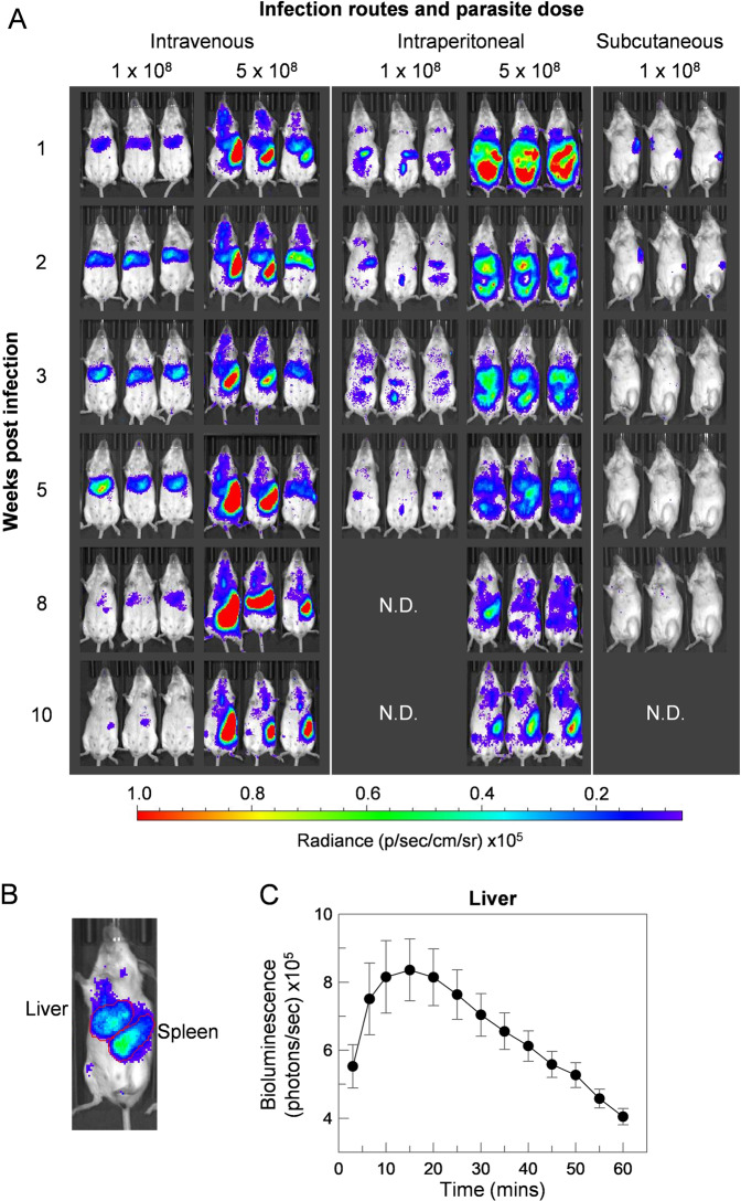
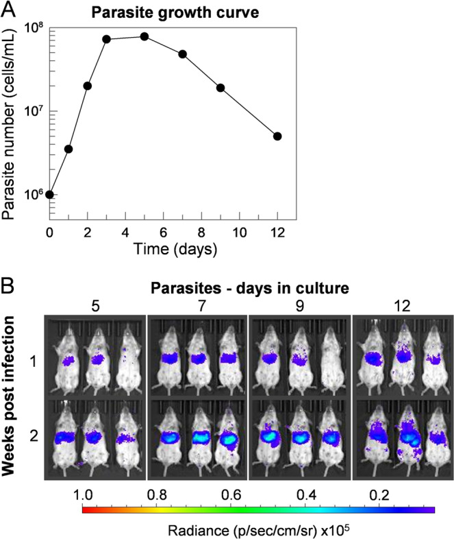
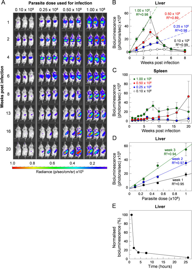
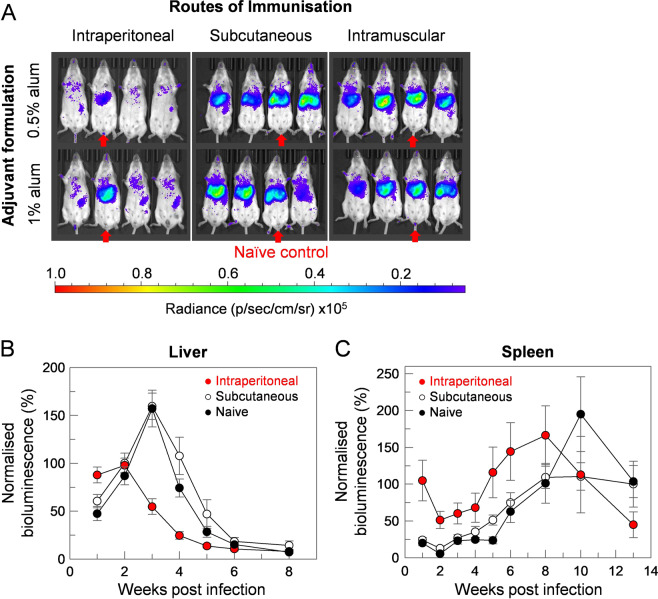
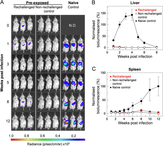
Visceral leishmaniasis is an infectious parasitic disease caused by the protozoan parasites Leishmania donovani and Leishmania infantum. The drugs currently used to treat visceral leishmaniasis suffer from toxicity and the emergence of parasite resistance, and so a better solution would be the development of an effective subunit vaccine; however, no approved vaccine currently exists. The comparative testing of a large number of vaccine candidates requires a quantitative and reproducible experimental murine infection model, but the parameters that influence infection pathology have not been systematically determined. To address this, we have established an infection model using a transgenic luciferase-expressing L. donovani parasite and longitudinally quantified the infections using in vivo bioluminescent imaging within individual mice. We examined the effects of varying the infection route, the site of adjuvant formulation administration, and standardised the parasite preparation and dose. We observed that the increase in parasite load within the liver during the first few weeks of infection was directly proportional to the parasite number in the initial inoculum. Finally, we show that immunity can be induced in pre-exposed animals that have resolved an initial infection. This murine infection model provides a platform for systematic subunit vaccine testing against visceral leishmaniasis.


Conflict of interest statement

The authors declare no competing interests.

Figures

Figure 1
Figure 1
The route of parasite administration in experimental infections of L. donovani influenced disease pathology. (A) Groups of five mice were infected with either day five (intraperitoneal and intravenous delivery) or day seven (subcutaneous) stationary-phase transgenic luciferase-expressing L. donovani promastigote cultures at the indicated doses. Parasite loads were quantified at the indicated times by administering the luciferase substrate, luciferin, and the animals imaged using an IVIS instrument. Three representative animals from the group are shown and are arranged so that the infection dynamics can be followed in each individual animal throughout the course of the infection. The route of parasite administration and dose influenced the progression of the infection. (B) Parasite loads were separately quantified in the liver or spleen by defining these organs as shown and measuring the associated bioluminescence. (C) Liver bioluminescence increased over time following luciferin injection, peaked at between 10 to 20 minutes before a steady decrease in signal over the next hour. Data points represent means ± S.E.M. (n = 5, from individual mice).
Figure 2
Figure 2
Infections are dependent on the duration of stationary phase parasite culture. (A) Fresh cultures of luciferase-expressing transgenic L. donovani parasites were established at an initial seeding density of 1 × 10⁶ cells/mL and growth was quantified over 12 days by counting live (motile) cells using a haemocytometer. After peaking at around days 3 to 7, motile parasite numbers declined. (B) Parasites were harvested at the specified time and used to infect groups of five mice intravenously. Infections were monitored during the initial 2–3 weeks by in vivo imaging. Three representative animals from the group are shown and are arranged so that the infection dynamics can be followed in each individual animal throughout the course of the infection.
Figure 3
Figure 3
The progression and severity of L. donovani infection of mice is dependent upon the initial inoculum size. (A) Groups of three mice were infected intravenously with different doses of day-seven stationary-phase L. donovani promastigotes and infections were monitored over 20 weeks by in vivo imaging. To quantify the parasite load, the bioluminescence (total flux, photon/sec) associated with the liver (B) and spleen (C) was measured. Mice were infected with 0.10 × 10⁸ (white circles), 0.25 × 10⁸ (blue circles), 0.50 × 10⁸ (red circles) or 1.00 × 10⁸ (green circles) parasites, respectively. Liver-associated bioluminescence increased linearly during the initial 1–3 weeks, analysed using linear regression in GraFit and plotted as dashed lines. The progression of infection of the liver during the initial three weeks was also analysed using linear regression and increases in bioluminescence were found to be directly proportional to the parasite dose for weeks 1 (black circles), 2 (blue circles) and 3 (green circles), respectively (D). The parasites’ rapid localisation and clearance in the liver within first 24 h of infection were determined in mice challenged with 1 × 10⁸ parasites (n = 5) (E). Liver bioluminescence was normalised using measurements at 1 hour post infection as control for 100%. Data points represent means ± S.E.M. (n = 3 for B, C and D) or (n = 5 for E).
Figure 4
Figure 4
Route of adjuvant formulation delivery affects disease pathology in a murine model of L. donovani infection. (A) Groups of five mice were immunised via intraperitoneal, subcutaneous or intramuscular routes of injection with a control protein expressed in a mammalian expression system adsorbed to 1% or 0.5% alum. Mice were challenged intravenously with 108 stationary-phase parasites, and an unimmunised mouse is present within each group (identified by red arrows) as a control. Infections were monitored by IVIS after 1 week and those immunised in the peritoneal cavity showed a reduction in parasite burden compared to the naïve control mice and those immunised by subcutaneous and intramuscular routes. (B,C) Groups of five mice were immunised via intraperitoneal or subcutaneous routes of injection with control protein adsorbed to 1% alum, challenged intravenously with 108 stationary-phase parasites and infections monitored compared to unimmunised naïve mice by in vivo imaging for 13 weeks. Bioluminescence (total flux, photon/sec) was quantified around defined regions of interest overlying the liver (B) and spleen (C) for naïve (black circles), subcutaneously-immunised (white circles) or intraperitoneally-immunised (red circles) animals. Data points represent means ± S.E.M. (n = 5). Relative bioluminescence was calculated by arbitrary normalisation to signals at week 2 (liver) and week 12 (spleen) of subcutaneously-immunised animals.
Figure 5
Figure 5
Mice that resolved an initial infection are resistant to reinfection with 108 parasites. (A) Two groups of five mice were pre-exposed to live parasites by intravenously infecting them with a suboptimal dose of 107 parasites per mouse; after 16 weeks, no parasites were detected by in vivo imaging. One group was then reinfected intravenously with 108 parasites (week 0), while the second group served as a control for any possible disease progression from the pre-exposure infection. A third group of naïve control mice was infected intravenously with 108 parasites as a positive control for virulence of the inoculum. By contrast to controls, mice that had been previously exposed to live parasites were immune to subsequent reinfection. Three representative animals from the group are shown and are arranged so that the infection dynamics can be followed in each individual animal throughout the course of the infection. (B,C) Bioluminesence (total flux, photon/sec) were quantified around defined regions of interest corresponding to the liver (B) and spleen (C). Group 1 (red circles, n = 4), Group 2 (white circles, n = 5) or Group 3 (black circles, n = 9) animals. Data points represent means ± S.E.M. of measurements. Relative bioluminescence was calculated by arbitrary normalisation to signals at week 2 (liver) and week 12 (spleen) of naive control-infected animals.

Similar articles

Cited by

References

    1. WHO. Leishmaniasis. https://wwwwhoint/news-room/fact-sheets/detail/leishmaniasis. (2019).
    1. González C, et al. Climate change and risk of leishmaniasis in north america: predictions from ecological niche models of vector and reservoir species. PLoS Negl. Trop. Dis. 2010;4:e585. doi: 10.1371/journal.pntd.0000585. - DOI - PMC - PubMed
    1. Purse BV, et al. How will climate change pathways and mitigation options alter incidence of vector-borne diseases? A framework for leishmaniasis in South and Meso-America. PLoS One. 2017;12:e0183583. doi: 10.1371/journal.pone.0183583. - DOI - PMC - PubMed
    1. Ready PD. Epidemiology of visceral leishmaniasis. Clin. Epidemiol. 2014;6:147–54. doi: 10.2147/CLEP.S44267. - DOI - PMC - PubMed
    1. Oryan A, Akbari M. Worldwide risk factors in leishmaniasis. Asian Pac. J. Tropical Medicine. 2016;9:925–32. doi: 10.1016/j.apjtm.2016.06.021. - DOI - PubMed

Publication types

MeSH terms

Substances