

Lumbar spinal stenosis and disc alterations affect the upper lumbar spine in adults with achondroplasia
- PMID: 32170149
- PMCID: PMC7070089
- DOI: 10.1038/s41598-020-61704-w
Lumbar spinal stenosis and disc alterations affect the upper lumbar spine in adults with achondroplasia
Abstract
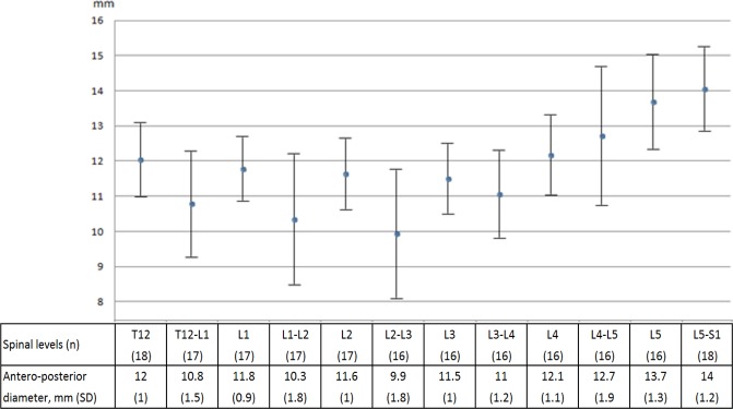
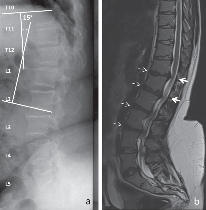
In achondroplasia, lumbar spinal stenosis arises from congenital dysplasia and acquired degenerative changes. We here aimed to describe the changes of the lumbar spinal canal and intervertebral disc in adults. We included 18 adults (age ≥ 18 years) with achondroplasia and lumbar spinal stenosis. Radiographs were used to analyze spinal-pelvic angles. Antero-posterior diameter of the spinal canal and the grade of disc degeneration were measured by MRI. Antero-posterior diameters of the spinal canal differed by spinal level (P < 0.05), with lower values observed at T12-L1, L1-2 and L2-3. Degrees of disc degeneration differed by intervertebral level, with higher degrees observed at L1-2, L2-3 and L3-4. A significant correlation was found between disc degeneration and thoraco-lumbar kyphosis at L2-3, between antero-posterior diameter of the spinal canal and lumbar lordosis at T12-L1 and L2-3, and between antero-posterior diameter of the spinal canal and thoraco-lumbar kyphosis at L1-2. Unlike the general population, spinal stenosis and disc degeneration involve the upper part of the lumbar spine in adults with achondroplasia, associated with thoraco-lumbar kyphosis and loss of lumbar lordosis.
Conflict of interest statement
The authors declare no competing interests.
Figures
References
MeSH terms
LinkOut - more resources
Full Text Sources
Medical
