. 2020 Mar 13;11(1):1361.
doi: 10.1038/s41467-020-15184-1.

How paired PSII-LHCII supercomplexes mediate the stacking of plant thylakoid membranes unveiled by structural mass-spectrometry

Affiliations

How paired PSII-LHCII supercomplexes mediate the stacking of plant thylakoid membranes unveiled by structural mass-spectrometry

Pascal Albanese et al. Nat Commun. .

Abstract

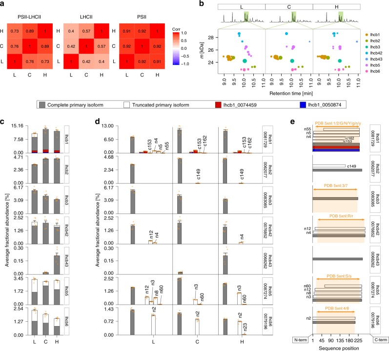
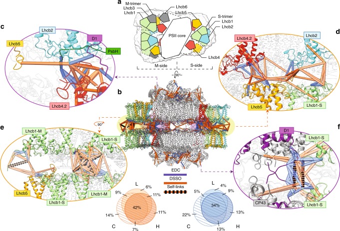
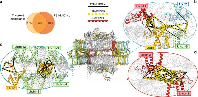
Grana are a characteristic feature of higher plants' thylakoid membranes, consisting of stacks of appressed membranes enriched in Photosystem II (PSII) and associated light-harvesting complex II (LHCII) proteins, together forming the PSII-LHCII supercomplex. Grana stacks undergo light-dependent structural changes, mainly by reorganizing the supramolecular structure of PSII-LHCII supercomplexes. LHCII is vital for grana formation, in which also PSII-LHCII supercomplexes are involved. By combining top-down and crosslinking mass spectrometry we uncover the spatial organization of paired PSII-LHCII supercomplexes within thylakoid membranes. The resulting model highlights a basic molecular mechanism whereby plants maintain grana stacking at changing light conditions. This mechanism relies on interactions between stroma-exposed N-terminal loops of LHCII trimers and Lhcb4 subunits facing each other in adjacent membranes. The combination of light-dependent LHCII N-terminal trimming and extensive N-terminal α-acetylation likely affects interactions between pairs of PSII-LHCII supercomplexes across the stromal gap, ultimately mediating membrane folding in grana stacks.


Conflict of interest statement

The authors declare no competing interests.

Figures

Fig. 1
Fig. 1. Workflow of the integrated MS-based approach for characterizing the light-driven modulation of paired PSII–LHCIIsc.
Isolation of heterogeneous mixtures of PSII–LHCIIsc from pea plants grown at different light intensities (L, low; C, moderate used as control; H, high). PSII–LHCIIsc preparations were used for TD-MS and XL-MS in vitro, performed with DSSO and EDC crosslinkers on two independently isolated PSII–LHCIIsc for each light condition pooled together. DSSO XL-MS in situ was conducted on thylakoid membranes isolated from three independent batches of pea plants grown in moderate (C) light.
Fig. 2
Fig. 2. TD-MS profiling of LHCII diversity.
a Spearman correlation of mass features detected in the PSII–LHCIIsc L, C and H samples for PSII–LHCIIsc (left), LHCII (middle), and PSII (right) proteins. b Region of the LC–MS chromatogram corresponding to the elution of LHCII proteoforms displayed as assigned mass features versus retention time (see Supplementary Fig. 1 for overview of all proteins detected in TD-MS). c Average abundances of LHCII proteins and d detailed average abundances of the distinct isoforms and proteoforms for each LHCII protein, accession number of the primary isoform is reported in the box on the right (see Supplementary Fig. 3 for quantification of distinct proteoforms detected in TD-MS). Error bars represent standard error of the mean abundance for each proteoform. e Schematic representation of sequence alignment of LHCII isoforms and proteoforms detected in TD-MS analyses and corresponding sequences resolved in the high-resolution structure of the PSII–LHCII supercomplex from pea plants (PDB: 5xnl, and chains therein; the orange box highlights the portion of protein sequence with resolved structure).
Fig. 3
Fig. 3. Mapping of crosslinks in the paired PSII–LHCIIsc predicted structural model.
a Schematic top-view of the (C2S2M)×2 fitted in the cryo-EM map EMD-3825, showing the overall arrangement of the PSII–LHCIIsc subunits in the predicted models 1–3 (M-side and S-side are indicated). b Side-view with mapped crosslinks within the distance cut-off of 17 Å for EDC (blue lines) and 33 Å for DSSO (orange lines), and DSSO self-links (orange-black dashed lines). Venn diagrams showing the overlap between datasets of PSII–LHCIIsc L, C and H samples; only crosslinks present in at least two out of three samples were considered. The enlarged views highlight the crosslinks involving Lhcb2 (c, d) and Lhcb1 (e, f) at the periphery of the supercomplex (d, e) and close to the PSII core (c, f). Subunits are coloured as follows: Lhcb1 in light green; Lhcb2 in cyan; Lhcb4.2 in red; Lhcb5 in yellow; D1 in purple, PsbH in green and CP43 in grey. Subunits not involved in crosslinks are left transparent.
Fig. 4
Fig. 4. Mapping of Lhcb4.2 crosslinks putatively responsible for the structural anchor of paired PSII–LHCIIsc.
a Side-view of the (C2S2M)x2 fitted in the cryo-EM map EMD-3825, with the “knot” connecting density highlighted in red. b Enlarged view highlighting the inter-protein crosslinks involving the N-terminus (Arg1–Asp27) and the long hairpin (Pro42–Phe87) of Lhcb4.2. These two regions of the N-terminal loop of the Lhcb4.2 are shown in black in the inset. Side-view c and end-view d of the putative site of interaction between the flexible N-termini (Arg1–Asp27) of two Lhcb4.2 subunits facing from adjacent supercomplexes are shown, and their ~18 Å displacement from the “knot” density is indicated. Crosslinks within the distance cut-off of 17 Å for EDC (blue lines) and of 33 Å for DSSO (orange lines), and DSSO self-links (orange-black dashed lines) are shown. Subunits are coloured as follows: Lhcb2 in cyan; Lhcb4.2 in red; PsbH in green and CP47 in grey. Subunits not involved in crosslinks are left transparent.
Fig. 5
Fig. 5. Detection of paired PSII–LHCIIsc in stacked thylakoid membranes by in situ XL-MS with DSSO.
a Side-view of the (C2S2M)×2 with mapped crosslinks found in at least two out of three thylakoid samples and two out of three PSII–LHCIIsc samples, together with the overlap of the two datasets. Enlarged views matching Figs. 3d, e and 4d are shown to highlight the positioning of Lhcb2 (b) and the occurrence of Lhcb1 and Lhcb4.2 mutual interactions (c, d) in the thylakoid membranes. Crosslinks within the distance cut-off of 33 Å for DSSO either for isolated PSII–LHCIIsc (black lines) or thylakoid membranes (yellow dashed lines), and DSSO self-links (red dashed lines) are shown. Subunits not involved in crosslinks are left transparent.
Fig. 6
Fig. 6. Map of acetylated N-terminal domains of PSII–LHCIIsc subunits spanning the stromal gap.
Schematic top- and side-view of the (C2S2M)×2, highlighting the acetylated N-terminal domains of the proteins spanning across the stromal gap detected and quantified by TD-MS (red box indicates acetylation rate of the complete primary isoform in any light condition, above 98% solid line; above 90% dashed line). Trimming position(s) and putative phosphorylation sites (based on homologous phosphosites previously detected in other plants) are indicated. N-α-acetylation (ac) detected in crosslinked peptides is shown. N-α-acetylation detected by XL-MS in vitro on isolated PSII–LHCIIsc (treated with DSSO, pentagon; treated with EDC, square) and in situ on the thylakoid membranes (treated with DSSO; triangle) is shown.

References

    1. Allen JP. Recent innovations in membrane-protein structural biology. F1000Research. 2019;8:211. doi: 10.12688/f1000research.16234.1. - DOI - PMC - PubMed
    1. Barber J. Photosystem II: the water-splitting enzyme of photosynthesis. Cold Spring Harb. Symp. Quant. Biol. 2012;77:295–307. doi: 10.1101/sqb.2012.77.014472. - DOI - PubMed
    1. Pagliano C, Saracco G, Barber J. Structural, functional and auxiliary proteins of photosystem II. Photosynth. Res. 2013;116:167–188. doi: 10.1007/s11120-013-9803-8. - DOI - PubMed
    1. Hankamer B, Morris EP, Barber J. Revealing the structure of the oxygen-evolving core dimer of photosystem II by cryoelectron crystallography. Nat. Struct. Biol. 1999;6:560–564. doi: 10.1038/9341. - DOI - PubMed
    1. Umena Y, Kawakami K, Shen J-R, Kamiya N. Crystal structure of oxygen-evolving photosystem II at a resolution of 1.9 Å. Nature. 2011;473:55–60. doi: 10.1038/nature09913. - DOI - PubMed

Publication types

MeSH terms

Substances