. 2020 May;80(5):e7-e13.
doi: 10.1016/j.jinf.2020.03.007. Epub 2020 Mar 21.

Clinical and CT imaging features of the COVID-19 pneumonia: Focus on pregnant women and children

Affiliations

Clinical and CT imaging features of the COVID-19 pneumonia: Focus on pregnant women and children

Huanhuan Liu et al. J Infect. 2020 May.

Abstract

Background: The ongoing outbreak of COVID-19 pneumonia is globally concerning. We aimed to investigate the clinical and CT features in the pregnant women and children with this disease, which have not been well reported.

Methods: Clinical and CT data of 59 patients with COVID-19 from January 27 to February 14, 2020 were retrospectively reviewed, including 14 laboratory-confirmed non-pregnant adults, 16 laboratory-confirmed and 25 clinically-diagnosed pregnant women, and 4 laboratory-confirmed children. The clinical and CT features were analyzed and compared.

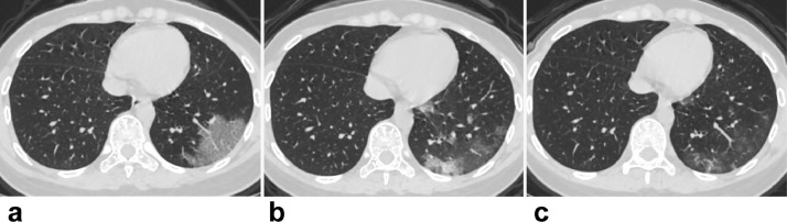
Findings: Compared with the non-pregnant adults group (n = 14), initial normal body temperature (9 [56%] and 16 [64%]), leukocytosis (8 [50%] and 9 [36%]) and elevated neutrophil ratio (14 [88%] and 20 [80%]), and lymphopenia (9 [56%] and 16 [64%]) were more common in the laboratory-confirmed (n = 16) and clinically-diagnosed (n = 25) pregnant groups. Totally 614 lesions were detected with predominantly peripheral and bilateral distributions in 54 (98%) and 37 (67%) patients, respectively. Pure ground-glass opacity (GGO) was the predominant presence in 94/131 (72%) lesions for the non-pregnant adults. Mixed consolidation and complete consolidation were more common in the laboratory-confirmed (70/161 [43%]) and clinically-diagnosed (153/322 [48%]) pregnant groups than 37/131 (28%) in the non-pregnant adults (P = 0·007, P < 0·001). GGO with reticulation was less common in 9/161 (6%) and 16/322 (5%) lesions for the two pregnant groups than 24/131 (18%) for the non-pregnant adults (P = 0·001, P < 0·001). The pulmonary involvement in children with COVID-19 was mild with a focal GGO or consolidation. Twenty-three patients underwent follow-up CT, revealing progression in 9/13 (69%) at 3 days whereas improvement in 8/10 (80%) at 6-9 days after initial CT scans.

Interpretation: Atypical clinical findings of pregnant women with COVID-19 could increase the difficulty in initial identification. Consolidation was more common in the pregnant groups. The clinically-diagnosed cases were vulnerable to more pulmonary involvement. CT was the modality of choice for early detection, severity assessment, and timely therapeutic effects evaluation for the cases with epidemic and clinical features of COVID-19 with or without laboratory confirmation. The exposure history and clinical symptoms were more helpful for screening in children versus chest CT.

Keywords: COVID-19 pneumonia; CT; Children; Pregnancy.


Figures

Fig. 1
Fig. 1
Chest CT images of a 29-year-old non-pregnant female with laboratory-confirmed COVID-19 pneumonia, presenting fever and cough for 2 days. (a) Axial thin-section CT showed the lesion was located in the peripheral region of left lower lobe, showing GGO with reticulation. (b) At the re-examined CT 6 days later, decreased size of the initial lesion and more GGOs with consolidation lesions were noted. (c) The second follow-up CT 7 days later, improvement with fewer lesions and decreased density were depicted.
Fig. 2
Fig. 2
Chest CT images of three pregnant women with laboratory-confirmed COVID-19 pneumonia. (a) 28-year-old female with 34 weeks plus 5 days pregnancy, presenting fever and loss of appetite for 2 days, GGO with consolidation in peripheral distribution was detected at CT in the left upper lobe. (b) 30-year-old female with 33 weeks pregnancy, presenting fever and fatigue for 4 days, GGO with consolidation and reticulation were identified with peripheral distribution in the right lower lobe. (C) 30-year-old female with 31 weeks pregnancy, presenting fever and cough for 2 days, complete consolidations were detected with peripheral distribution in the lower lobes of bilateral lungs.
Fig. 3
Fig. 3
Chest CT images of a 28-year-old female with 36 weeks plus 1 day pregnancy, with clinically-diagnosed COVID-19 pneumonia, presented post-partum low grade fever. (a) Baseline CT imaging demonstrated typical CT features of COVID-19 pneumonia, with multiple GGOs and GGOs with reticulation lesions predominantly peripheral distribution in both lungs. (b) Follow-up CT 10 days later after antiviral therapy, demonstrated improvement with fewer lesions and few consolidation lesions left.
Fig. 4
Fig. 4
Positive chest CT of 3 children with laboratory confirmation and atypical CT features of COVID-19 pneumonia. (a) 11-month-old child with fever, cough, and fatigue for 6 days. The only consolidation lesion was located in central region of the right lower lobe. (b) 9-year-old child with fever for 3 days. The only GGO lesion was located in the interlobular subpleural area of right middle lobe. (c) 2-month-old child with cough for 2 days and the respiratory syncytial virus positive, one of the multiple consolidation lesions with air bronchogram was located in the right upper lobe, and pleural effusion was also noted.

Comment in

  • Unfavorable outcomes in pregnant patients with COVID-19.
    Huang W, Zhao Z, He Z, Liu S, Wu Q, Zhang X, Qiu X, Yuan H, Yang K, Tang X, Zhang S. Huang W, et al. J Infect. 2020 Aug;81(2):e99-e101. doi: 10.1016/j.jinf.2020.05.014. Epub 2020 May 15. J Infect. 2020. PMID: 32417313 Free PMC article. No abstract available.

References

    1. Zhu N, Zhang D, Wang W, Li X, Yang B, Song J. A Novel Coronavirus from Patients with Pneumonia in China, 2019. N Engl J Med. 2020;382(8):727–733. - PMC - PubMed
    1. Huang C, Wang Y, Li X, Ren L, Zhao J, Hu Y. Clinical features of patients infected with 2019 novel coronavirus in Wuhan. China. Lancet. 2020;395(10223):497–506. - PMC - PubMed
    1. Chen N, Zhou M, Dong X, Qu J, Gong F, Han Y. Epidemiological and clinical characteristics of 99 cases of 2019 novel coronavirus pneumonia in Wuhan, China: a descriptive study. Lancet. 2020;395(10223):507–513. - PMC - PubMed
    1. The International Committee on Taxonomy of Viruses (ICTV) Coronaviridae Study Group. Naming the 2019 Coronavirus. https://talk.ictvonline.org/.
    1. Chan JF, Yuan S, Kok KH, To KK, Chu H, Yang J, et al. A familial cluster of pneumonia associated with the 2019 novel coronavirus indicating person-to-person transmission: a study of a family cluster. 2020;395(10223):514-523. - PMC - PubMed

MeSH terms