Review
. 2020 Jul;1865(7):158681.
doi: 10.1016/j.bbalip.2020.158681. Epub 2020 Mar 12.

S1P and plasmalogen derived fatty aldehydes in cellular signaling and functions

Affiliations
Review

S1P and plasmalogen derived fatty aldehydes in cellular signaling and functions

David L Ebenezer et al. Biochim Biophys Acta Mol Cell Biol Lipids. 2020 Jul.

Abstract

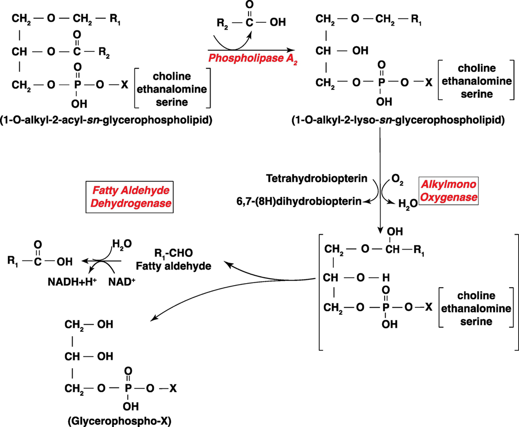
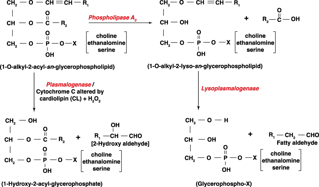
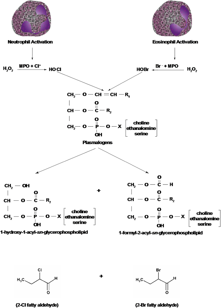
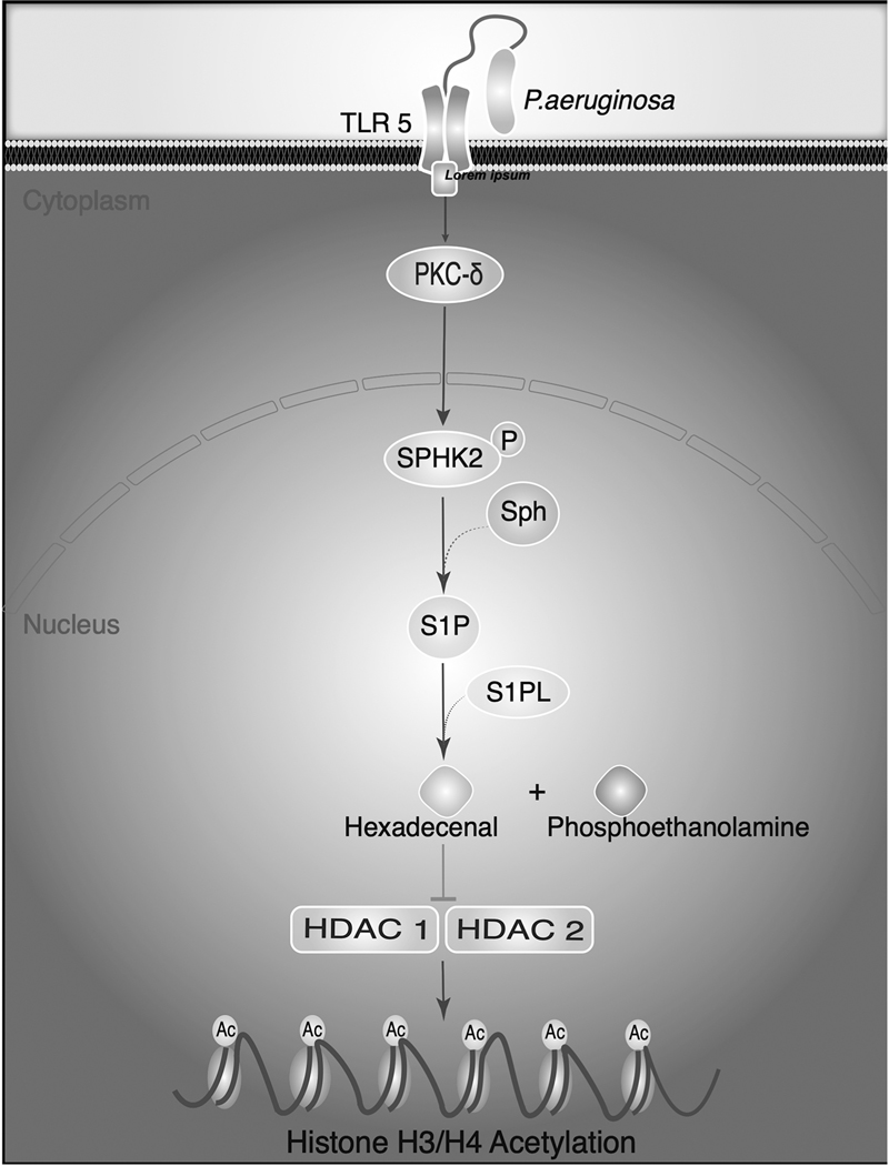
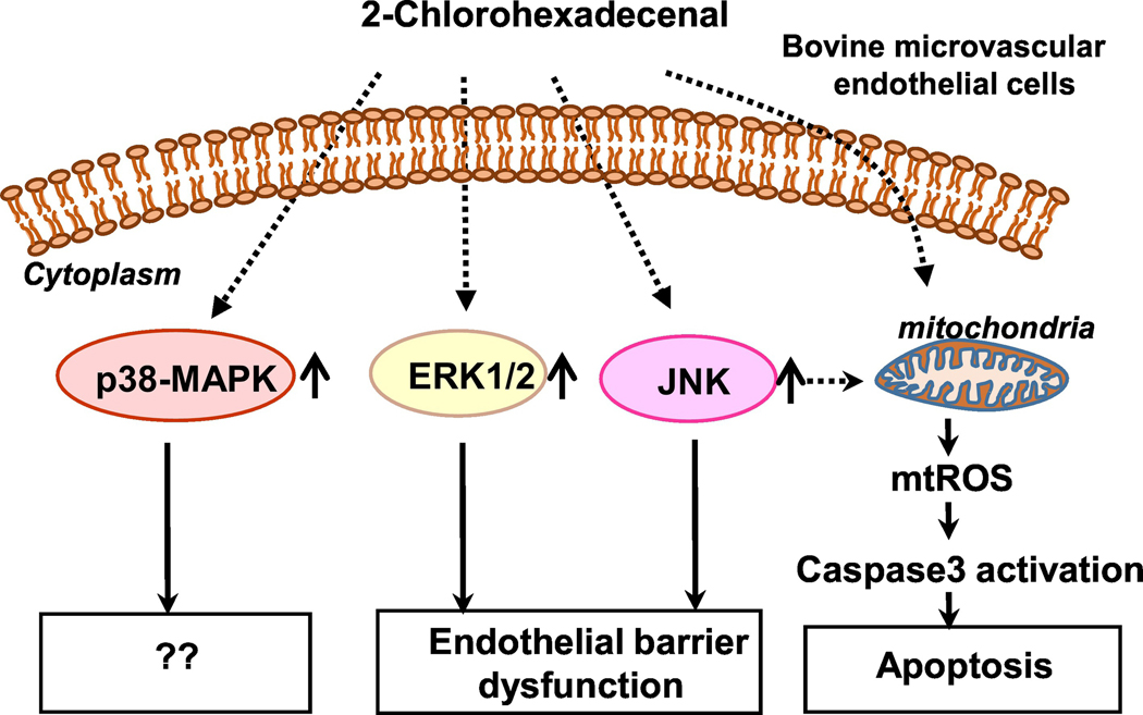
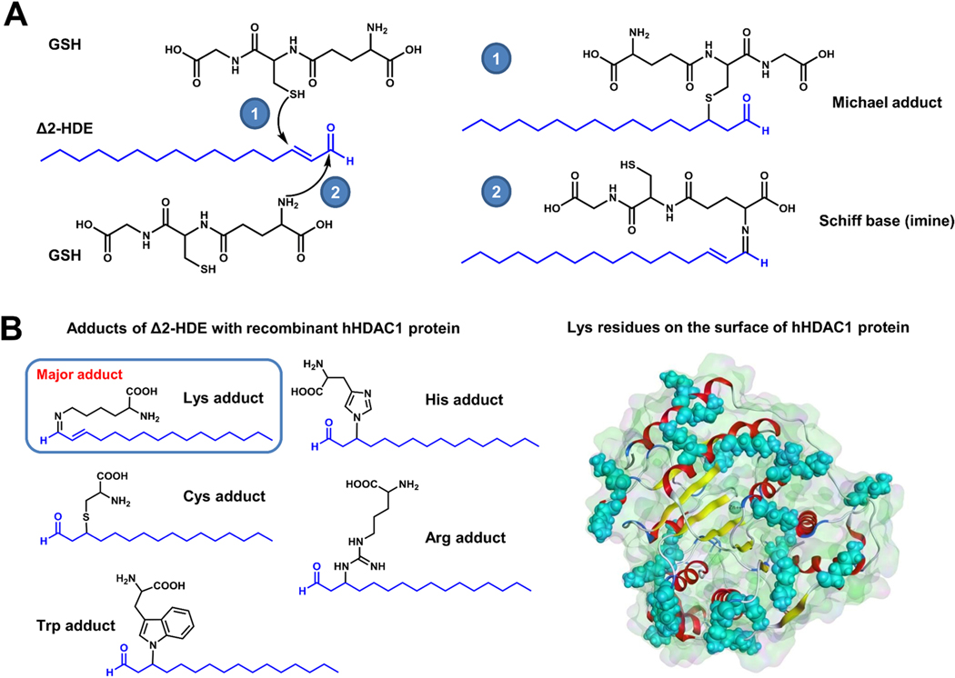
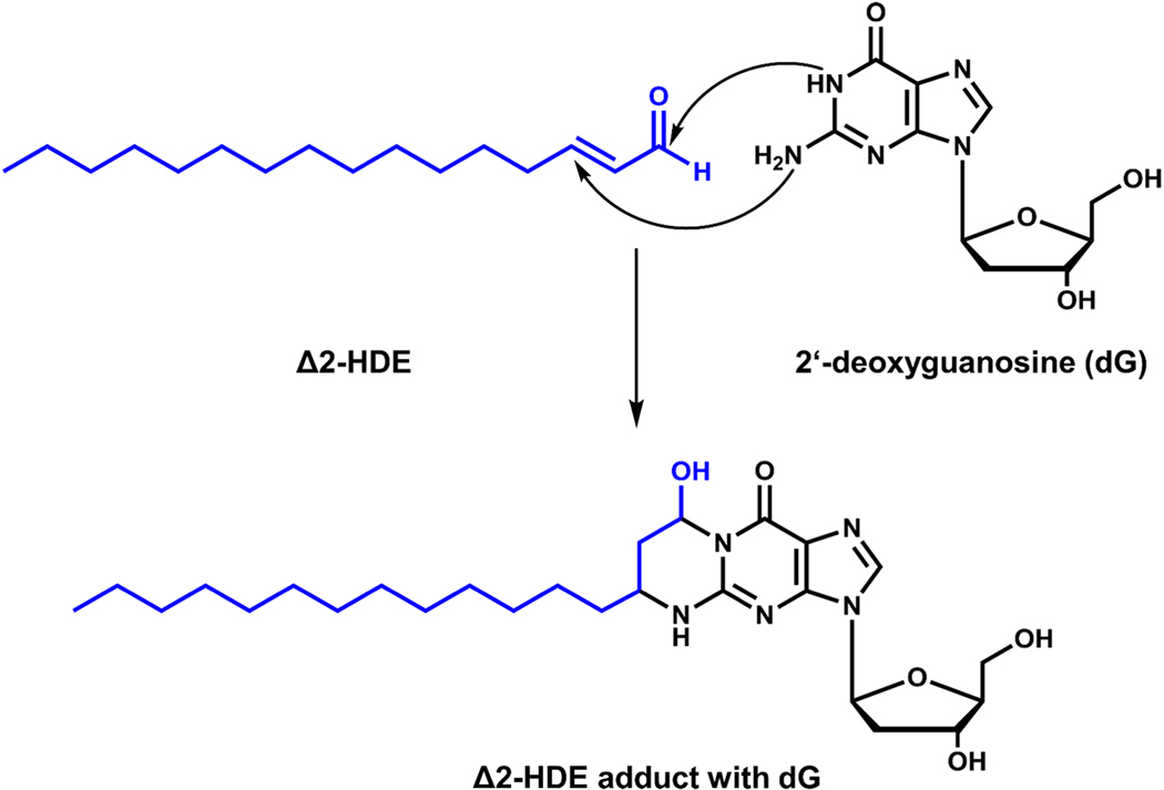
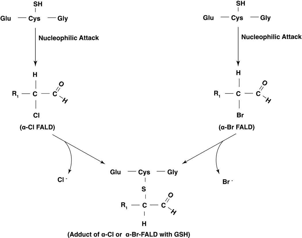
Long-chain fatty aldehydes are present in low concentrations in mammalian cells and serve as intermediates in the interconversion between fatty acids and fatty alcohols. The long-chain fatty aldehydes are generated by enzymatic hydrolysis of 1-alkyl-, and 1-alkenyl-glycerophospholipids by alkylglycerol monooxygenase, plasmalogenase or lysoplasmalogenase while hydrolysis of sphingosine-1-phosphate (S1P) by S1P lyase generates trans ∆2-hexadecenal (∆2-HDE). Additionally, 2-chloro-, and 2-bromo- fatty aldehydes are produced from plasmalogens or lysoplasmalogens by hypochlorous, and hypobromous acid generated by activated neutrophils and eosinophils, respectively while 2-iodofatty aldehydes are produced by excess iodine in thyroid glands. The 2-halofatty aldehydes and ∆2-HDE activated JNK signaling, BAX, cytoskeletal reorganization and apoptosis in mammalian cells. Further, 2-chloro- and 2-bromo-fatty aldehydes formed GSH and protein adducts while ∆2-HDE formed adducts with GSH, deoxyguanosine in DNA and proteins such as HDAC1 in vitro. ∆2-HDE also modulated HDAC activity and stimulated H3 and H4 histone acetylation in vitro with lung epithelial cell nuclear preparations. The α-halo fatty aldehydes elicited endothelial dysfunction, cellular toxicity and tissue damage. Taken together, these investigations suggest a new role for long-chain fatty aldehydes as signaling lipids, ability to form adducts with GSH, proteins such as HDACs and regulate cellular functions.

Keywords: Hexadecenal; Long-chain fatty aldehydes; Lysoplasmalogenase; Plasmalogenase; S1P; S1P Lyase.


Conflict of interest statement

Declaration of competing interest The authors declare no conflict of interest and no financial obligations.

Figures

Figure 1:
Figure 1:. Pathways involved in the degradation of phospholipids and sphingolipids.
(A) Phospholipases mediated hydrolysis of 1,2-diacyl-sn-glycero-3-phosphocholine (PC) by phospholipases and lysophospholipase D (lyso PLD) or autotaxin (ATX). Hydrolysis of 1,2-diacyl-sn-glycero-3-phosphocholine (PC) by phospholipase (PL) A1, or A2, releases fatty acid from carbon 1 or carbon 2 of PC and generates 2-lyso-1-acyl- or 1-lyso-2-acyl-sn-glycero-3phosphocholine (LPC). Hydrolysis of PC by PLC generates diacylglycerol (DAG) and phosphocholine. DAG kinase (DAGK) converts DAG to 1,2-diacyl-sn-glycero-3-phosphate (PA), which is acted by PA specific PLA1 or PLA2 to give rise to 2-acyl- or 1-acyl-sn-glycero-3-phosphate (LPA). PLD catalyzes the hydrolysis of PC to PA and choline. 2-lyso-1-acyl- or 1-lyso-2-acyl-sn-glycero-3-phosphocholine (LPC) generated by PLA1 or PLA2 can be hydrolyzed by lyso PLD or ATX to 2-acyl- or 1-acyl-sn-glycero-3-phosphate (LPA). PLA2 mediated hydrolysis of PC releases polyunsaturated fatty acids C20:4, C22:5 and C22:6 from carbon 2, which are subsequently converted to prostanoids, leukotrienes, hydroperoxyeicosatetraenoic acids (HETES) and hydroperoxyeicosatetraenoic acids (HPETES) mediated by cyclooxygenases, lipoxygenases, peroxidases and dehydrogenases. (B) Hydrolysis of sphingomyelin by sphingomyelinase, ceramidase and S1P lyase. Sphingomyelin (SM) is hydrolyzed by sphingomyelinase (SMase) to produce ceramide that is acted by ceramidase(s) to generate sphingosine. Sphingosine is converted to sphingosine-1-phosphate (S1P) by sphingosine kinases (SPHKs) 1 and 2. Sphingosine-1-phosphate, thus generated is converted back to sphingosine by sphingosine-1-phosphate phosphatases (SPPs) and lipid phosphate phosphatases (LPPs) or to trans Δ2-hexadecenal and ethanolamine phosphate by S1P lyase, a pyridoxal phosphate-dependent enzyme.
Figure 2:
Figure 2:. Pathway of long-chain fatty aldehyde production from 1-O- alkyl phospholipids by phospholipase A2 and alkylmono-oxygenase.
Phospholipase A2 dependent hydrolysis of 1-O-alkyl-2-acyl-sn-glycerophospholipid generates 1-O-alkyl-2-lyso-sn-glycerophospholipid (Alkyl- lysophospholipid). An alkylmono-oxygenase converts the alkyl-lysophospholipid to a long-chain fatty aldehyde, R1─CHO that gives rise to R1─COOH catalyzed by fatty aldehyde dehydrogenase. X denotes the polar head group, which could be choline, ethanolamine or serine.
Figure 3:
Figure 3:. Pathways of plasmalogenase and lysoplasmalogenase in generating long-chain fatty aldehydes from 1-O-alkenyl phospholipids.
Plasmalogens are glycerophospholipids that have a 1-O-alkenyl- group linked to carbon 1 atom of the phospholipid, and lysoplasmalogens are derived from plasmalogens by the action of phospholipase A2. Plasmalogenase is cytochrome c that is activated by a cardiolipin (CL) and hydrogen peroxide (H2O2) in the mitochondria and releases 2-hydroxyl aldehyde and 1-lyso-2-acyl-sn-glycerophospholipid. Lysoplasmalogenase hydrolyzes 1-O-alkenyl-2-lyso-sn-glycerophospholipid generated from 1-O-alkenyl-2-acyl-sn-glycerophospholipids by PLA2 to fatty aldehyde and water soluble glycerophospho-X where X is choline, ethanolamine or serine.
Figure 4:
Figure 4:. Pathways of generation of 2-chloro- and 2-bromo-fatty aldehydes from plasmalogens by hypochlorous and hypobromous acids.
Activation of neutrophils and eosinophils generates hypochlorous acid (HOCl) and hypobromous acid (HOBr), respectively. HOCl and HOBr by a non-enzymatic mechanism attacks the –CH=CH_ bond of plasmalogens generating 2-chloro- or 2-bromo- fatty aldehydes and 1-hydroxy (lyso)-2-acyl- and 1-formyl-2-acyl-sn-glycerophospholipid where X is choline, ethanolamine or serine. Neutrophil activation releases H2O2, which in the presence of myeloperoxidase (MPO) and chloride ion produces HOCl. Activation of eosinophils releases H2O2 plus eosinophil peroxidase that reacts with bromide ion to form HOBr.
Figure 5:
Figure 5:. Degradation of sphingosine-1-phosphate (S1P).
Sphingosine-1-phosphate (S1P) generated from sphingosine by sphingosine kinases 1 and 2 (SPHK1 & 2) is irreversibly metabolized by S1P lyase, a pyridoxal phosphate dependent enzyme, to trans-Δ2-hexadecenal (Δ2-HDE) and ethanolamine phosphate. Δ2-HDE is further oxidized by fatty aldehyde dehydrogenase (FALDH) followed by coupling to coenzyme A (CoA) by acyl-CoA synthase. The product hexadecenoyl-CoA can be saturated by means of trans-2-enoyl-CoA reductase to form palmitoyl-CoA that can serve as building block for glycerolipid (and sphingolipid) synthesis.
Figure 6.
Figure 6.. Trans Δ2-hexadecenal stimulates signaling pathways in HEK293 and C6 glioma cells.
(A) Trans-Δ2-hexadecenal (Δ2-HDE) stimulates cytoskeletal reorganization and apoptosis in HEK293/NIH3T3/HeLa cells via activation of MKK4/MKK7►MLK3►JNK signaling. Trans Δ2-HDE-induced apoptosis involved Jun N-terminal kinase (JNK) phosphorylation that was reactive oxygen species (ROS) dependent accompanied by Bax activation, translocation of Bax and Bim to mitochondria, and cytochrome release (104). (B) Exogenous addition of trans Δ2-HDE to C6 glioma cells activated p38-mitogen activated protein kinase (p38-MAPK), extracellular signal-related kinase (ERK) 1/2) and phosphatidylinositol 3-kinase (PI3K) signaling pathways that regulated glioma cell proliferation (105).
Figure 7:
Figure 7:. Nuclear S1P lyase generated trans Δ2-hexadecenal from nuclear S1P modulates HDAC activity and histone acetylation.
Pseudomonas aeruginosa infection of mouse lung and lung epithelial cells in vitro stimulates phosphorylation of sphingosine kinase 2 (SPHK2) in the cytoplasm mediated by protein kinase C (PKC)-δ. Activated SPHK2 is translocated to the nucleus of the epithelial cell where it converts sphingosine to sphingosine-1-phosphate (S1P). Although S1P lyase is predominantly localized in the endoplasmic reticulum (ER), presence of S1P lyase in the nuclear preparations was detected by Western blotting and purity of the nuclear preparations from ER was verified by electron microscopy and immunostaining of the preparations with specific markers for ER, Golgi, cytoplasm and nuclear membrane. S1P generated in the nucleus by nuclear SPHK2 is hydrolyzed by S1P lyase to generate trans Δ2-hexadecenal (Δ2-HDE) and ethanolamine phosphate. S1P or Δ2-HDE generated from S1P modulates HDAC1/2 activity and H3/H4 histone acetylation pattern in lung epithelial cells (85, 86).
Figure 8:
Figure 8:. 2-Chlorohexadecanal signaling and endothelial barrier dysfunction.
Exogenous addition of 2-chlorohexadecanal (2-ClHDA) to brain microvascular endothelial cells stimulated extracellular signal-related kinase1/2 (ERK1/2), p38 mitogen-activated protein kinase (p38- MAPK) and Jun N-terminal kinase (JNK) signaling pathways and blocking ERK1/2 and JNK, but not p38 MAPK, attenuated 2-ClHDA-mediated endothelial dysfunction. 2-ClHDA also caused induced mitochondrial dysfunction including increased mitochondrial reactive oxygen species (mtROS), and apoptosis via activation of caspase 3.
Figure 9:
Figure 9:. Trans-Δ2-hexadecenal forms adducts with glutathione, and HDAC1 in vitro.
(A) Incubation of Δ2-HDE with glutathione (GSH) in vitro resulted in formation of Michael adducts (1), and Schiff’s base adducts (2) as determined by LC-MS/MS. (B) Incubation of Δ2-HDE with recombinant hHDAC1 in vitro generated five different adducts (left panel) with the Schiff’s base (imine) of lysine and Δ2-HDE by far was the most predominant species generated as detected by LC-MS/MS. This observation is consistent with the high amount of Lys residues in the sequence of hHDAC1 (40x) and their exposed localization on the protein’s surface (shown in cyan, right panel).
Figure 10:
Figure 10:. Adduct formation between trans-Δ2-hexadecenal and 2’-deoxyguanosine.
Scheme depicts the adduct formation between trans-Δ2-hexadecenal (Δ2-HDE) and DNA nucleoside 2’-deoxyguanosine (dG) in vitro as detected by LC-MS/MS.
Figure 11:
Figure 11:. Glutathione adducts of 2-chlorofatty aldehydes and 2-bromofatty aldehydes.
2-Chlorofatty aldehydes (2-ClFALDs) and 2-bromofatty aldehydes (2-BrFALDs) generated by hypochlorous acid (HOCl) or hypobromous acid (HOBr) react with a nucleophile such as glutathione (GSH) and form FALD-GSH adducts in vitro and in animals exposed to either chlorine or bromine gas.

Similar articles

Cited by

References

    1. O’Donnell VB, Rossjohn J, Wakelam MJ, Phospholipid signaling in innate immune cells, J Clin Invest, 128 (2018) 2670–2679. - PMC - PubMed
    1. Aloulou A, Rahier R, Arhab Y, Noiriel A, Abousalham A, Phospholipases: An Overview, Methods Mol. Biol, 1835 (2018) 69–105. - PubMed
    1. Chap H, Forty five years with membrane phospholipids, phospholipases and lipid mediators: A historical perspective, Biochimie, 125 (2016) 234–249. - PubMed
    1. Eyster KM, The membrane and lipids as integral participants in signal transduction: lipid signal transduction for the non-lipid biochemist, Adv Physiol Educ, 31 (2007) 5–16. - PubMed
    1. Phillips MC, Is ABCA1 a lipid transfer protein? J. Lipid Res, 59 (2018) 749–763. - PMC - PubMed

Publication types