. 2020 Jul;154(1):55-75.
doi: 10.1007/s00418-020-01861-1. Epub 2020 Mar 14.

Plasma cells, plasmablasts, and AID+/CD30+ B lymphoblasts inside and outside germinal centres: details of the basal light zone and the outer zone in human palatine tonsils

Affiliations

Plasma cells, plasmablasts, and AID+/CD30+ B lymphoblasts inside and outside germinal centres: details of the basal light zone and the outer zone in human palatine tonsils

Birte S Steiniger et al. Histochem Cell Biol. 2020 Jul.

Abstract

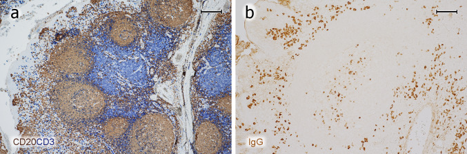
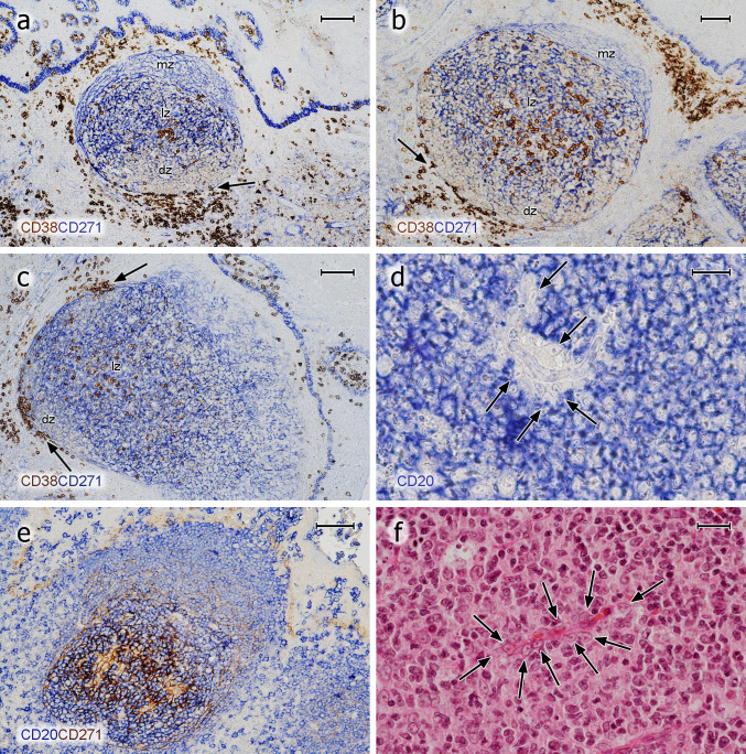
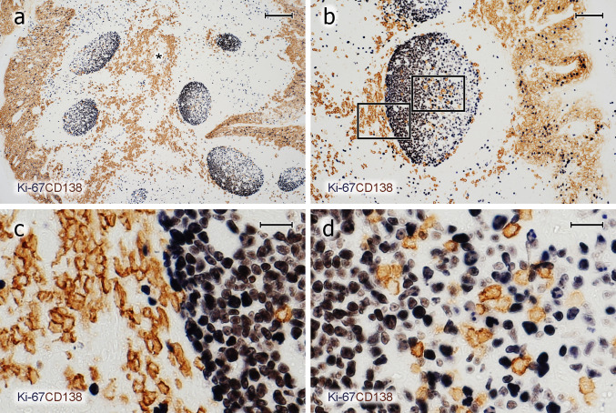
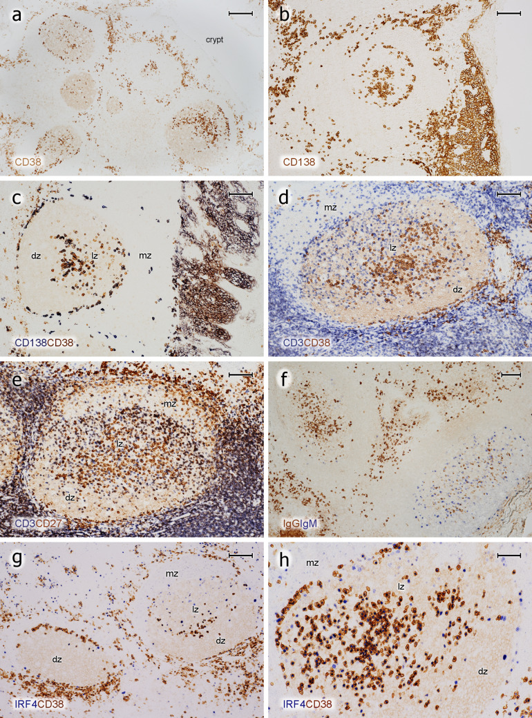
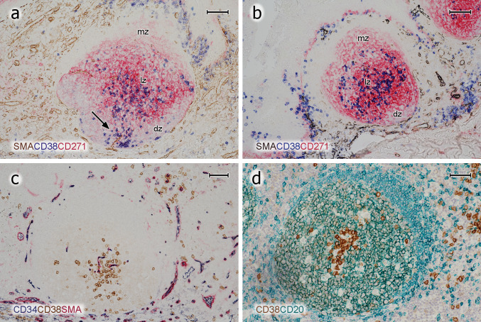
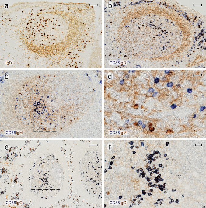
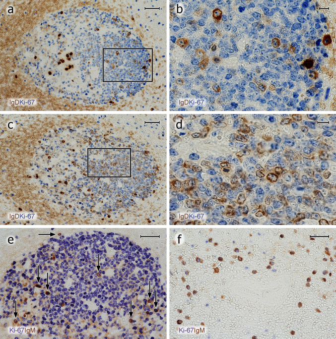
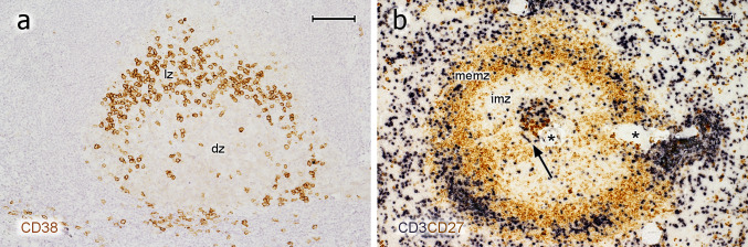
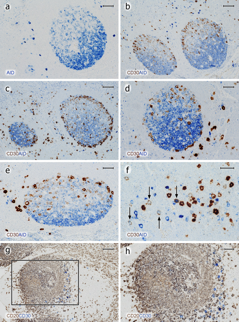
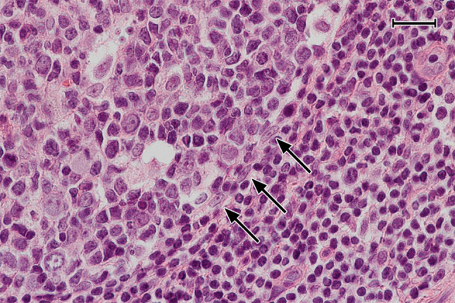
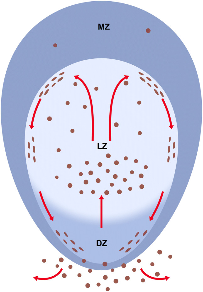
Plasma cells (PCs) in human palatine tonsils are predominantly located in the germinal centres (GCs), in the subepithelial space and near the deep connective tissue septa surrounding each crypt. We analysed the location, phenotype, and proliferation of GC PCs by immunohistology comparing them to PCs in the other two locations. Most PCs in GCs were strongly positive for CD38, CD138, CD27, IRF4, and intracellular (ic) IgG. They often accumulated in the basal light zone, but could also be found scattered in the entire light zone. In addition, rows of PCs occurred at the surface of the GC bordering the mantle zone, i.e., in the outer zone, and at the surface of the dark zone. The latter cells were often continuous with PCs in the extrafollicular area. The vast majority of GC PCs were negative for Ki-67. Only a few Ki-67+ plasmablasts, predominantly icIgG+ or icIgM+, were found inside GCs. In certain GCs PCs accumulated around capillaries and the adjacent perikarya of follicular dendritic cells (FDCs). Newly formed PCs might migrate from the basal to the superficial part of the light zone and then back to the dark zone surface to leave the GC. This guarantees an even distribution of secreted Ig for exchange with immune complexes on FDCs. The surface of the dark zone may also be an exit site for Ki-67+CD30+ B lymphoblasts, which seed perifollicular and extrafollicular sites. We speculate that these cells tend to downmodulate CD20 and activation-induced deaminase and further up-regulate CD30 when developing into pre-plasmablasts.

Keywords: B lymphoblasts; Germinal centres; Palatine tonsils; Plasma cells; Plasmablasts.


Conflict of interest statement

The authors are not aware of any conflict of interest.

Figures

Fig. 1
Fig. 1
Overview of T- and B-cell regions and distribution of PCs in a representative tonsil. a Staining for CD20 (mAb L26) in brown followed by visualisation of CD3 (polyclonal Abs) in blue colour. A crypt is located to the left. The connective tissue septum (unstained) in the right part of the image separates the crypt and adjacent lymphatic tissue from the lymphatic tissue associated with the next crypt to the right. The follicles are surrounded by a small perifollicular region with mixed T and B cells. The T-cell zone proper (blue region) is located deeper in the lamina propria in proximity to the connective tissue septum. b IcIgG+ PCs are primarily located beneath the crypt epithelium (left), in a GC (center), and near the connective septum between two crypts containing a vein (right). a, b 38-year-old female. Scale bar 200 µm (a, b)
Fig. 2
Fig. 2
Distribution of CD38++ PCs and pericapillary FDCs in tonsil GCs. ac Staining for CD38++ PCs (mAb SPC32, brown) followed by visualisation of CD271+ FDCs (mAb EP1039Y, blue). PCs may accumulate in the basal light zone and/or distribute evenly in the light zone and occur at the surface of the GC. They also form rows of cells at the dark zone surface, which are connected to PC clusters in the lamina propria outside the GC (arrows). The basal layer of the crypt epithelium is also CD271+. d FDCs at the surface of intra-GC microvessels (arrows) prevent CD20+ B lymphocytes (mAb L26, blue) from approaching the endothelium. e The unstained cells in d are CD271+, when CD20 is demonstrated in blue (mAb L26) followed by CD271 in brown (mAb EP1039Y). f The nuclei of the pericapillary FDCs (arrows) are also visible in routine HE-stained sections. mz mantle zone, lz light zone of GC, dz dark zone of GC. a, b 21-year-old female. c 12-year-old female. d, e 38-year-old female. f 57-year-old male. Scale bars 100 µm (a, c), 70 µm (b, e), 20 µm (d, f)
Fig. 3
Fig. 3
PCs inside and outside GCs do not proliferate. ad Visualisation of Ki-67 (mAb MIB-5, blue-black) followed by staining for CD138 (mAb BB-4, brown) shows that the vast majority of PCs inside and outside GCs is Ki-67. c, d Magnifications of the areas indicated in b. The entire crypt epithelium is also CD138+ (a, b). It covers both sides of the specimen in a and the right side in b. The brown cells in the central part of a represent PCs accompanying the central connective tissue septum (asterisk) between two crypts. ad 21-year-old female. Scale bars 200 µm (a), 100 µm (b), and 20 µm (c, d)
Fig. 4
Fig. 4
Phenotype of PCs in tonsil GCs. a Single staining for CD38 (mAb SPC32) and b CD138 (mAb BB-4). The crypt epithelium is located at the left and right margins in a and at the right margin in b. c Staining for CD138 (blue-black) followed by staining for CD38 (brown). d Staining for CD3 (polyclonal Abs, blue) followed by visualisation of CD38 (brown). e Visualisation of CD3 (dark blue) and subsequently of CD27 (mAb 137B4, brown). CD27++ PCs are located beneath the epithelium (upper rim of image) and in the GC. The superficial CD27+ cells in the mantle zone adjacent to the subepithelial PCs represent memory B cells. f Double staining for IgG (mAb RWP49, brown) and IgM (polyclonal Abs, blue). Two GCs differ in the prevalence of IgG+ (left GC) and IgM+ (right GC) PCs. The crypt epithelium and subepithelial PCs are hit in the centre and lower left corner of the image, where detritus in a crypt stains dark brown. g Overview of two follicles stained for IRF4 (mAb EP190, blue-black) and CD38 (mAb SPC32, brown) (h). Additional follicle stained in the same way showing a combination of the PC patterns of the two GCs in g. The epithelium is located near the upper or upper left parts of the image in d, e, g, h. mz mantle zone, lz light zone of GC, dz dark zone of GC. ah 21-year-old female. Scale bars = 200 µm (a), 100 µm (bg), 50 µm (h)
Fig. 5
Fig. 5
Localisation of PCs, FDCs, and capillaries in GCs. a Follicle stained for SMA (polyclonal Abs, brown), CD38 (blue), and CD271 (red). In this exceptional GC, CD38++ PCs also appear in the dark zone and accompany a microvessel (arrow). b Follicle stained for SMA (brown-black), CD38 (blue), and CD271 (red). CD38++ PCs are preferentially located in the basal light zone, where the density of red CD271+ FDC processes is highest. c Staining for CD34 (mAb QBend-10, blue), CD38 (mAb SPC32, brown), and SMA (red). CD38++ PCs often occur loosely distributed in the vicinity of CD34+ GC capillary endothelia in the light zone. d They may, however, also be directly adjacent to capillaries or perivascular FDCs (CD38 first stained in brown and, subsequently, CD20 in green). mz mantle zone, lz light zone of GC, dz dark zone of GC. a, d 57-year-old male. b, c 38-year-old female. Scale bars 70 µm (ac), 100 µm (d)
Fig. 6
Fig. 6
Expression of CD38 by icIg+ PCs in GCs. a Distribution of surface and icIgD in an icIgD+ GC. A minority of GCs contain a large number of IgD+ + PCs and also weakly IgD +  cells (cell surface or intracellular staining). The mantle zone (brown, right) is formed by small recirculating surface IgD+  B-lymphocytes. b GC stained first for CD38 (mAb SPC32, blue) and then for IgD (polyclonal Abs, brown). The dark blue PCs may be single or double positive. The entire GC is occupied by weakly IgD+ cells. These cells are less intensely stained than the surface IgD+ cells in the mantle zone (right). c Visualisation of CD38 in blue and icIgM in brown. The mantle zone is directed towards the left part of the image. d Higher magnification of area marked in c. There are several icIgM+ cells with low or absent staining for CD38. Weak surface staining of IgM+ mantle zone cells and staining of IgM+ immune complexes on FDCs is accepted in c and d to increase the sensitivity of the method. e In contrast to icIgM, staining for CD38 (mAb SPC32, blue-black) and icIgG (mAb RWP49, brown) reveals only very few icIgG+CD38± cells in this specimen. The right part of the GC contains the dark zone. f Higher magnification of area indicated in e. The crypt epithelium is located near the upper margin or upper right margin of the images in ac and near the left margin in e. af 38-year-old female. Scale bars 70 µm (ac), 14 µm (d), 100 µm (e), and 20 µm (f)
Fig. 7
Fig. 7
Proliferation of icIg+ cells. ad Visualisation of IgD+ cells (polyclonal Abs, brown) followed by detection of Ki-67 (mAb MIB-5, blue). a, b There are several Ki-67+icIgD+ plasmablasts in the dark zone and Ki67icIgD++ PCs in the basal light zone. The icIg+ cells in the dark zone do not exhibit the typical nuclear morphology of PCs. c, d In another follicle of the same individual, even more weakly IgD+ cells with Ki-67 expression are seen in the dark zone. It is impossible to distinguish intracellular and surface staining. Such GCs are infrequent. e Several dark zone Ki-67+ cells stained blue in the first step show icIgM in the second step (brown) of staining (vertical arrows). Single double-positive cells always occur at the surface of the dark zone (horizontal arrow). f In the outer T-cell region of a human spleen Ki-67 (blue) is demonstrated in about 50% of icIgM+ migratory plasmablasts (brown). The unstained structure in the centre of the image is a bending central artery. Same method as in e. The crypt epithelium is located beyond the left margin in a, c and below the bottom of the image in e. ae 38-year-old female. f 17-year-old male. Scale bars 50 µm (a, c), 10 µm (b, d), and 40 µm (e, f)
Fig. 8
Fig. 8
PCs in lymph node and spleen GCs. a Single staining for CD38 in a follicle of a cervical lymph node. The distribution of CD38++ PCs is similar to that in tonsils (compare Fig. 4a). lz light zone of GC, dz dark zone of GC. The mantle zone cannot be delineated. b Double staining for CD3 (dark blue) and CD27 (brown) in an adult human spleen. This method is optimal to visualise all regions of typical degenerated secondary follicles in normal human spleens (Steiniger et al. 2005, 2006). The follicle consists of a very small GC populated by CD27++ PCs and T cells (arrow), a broad inner mantle zone (imz, unstained), and a CD27+ memory B-cell zone (memz), which consists of the outer mantle zone and a superficial zone (not visualised in detail). Major numbers of T cells also occur in the superficial zone. Asterisks indicate a follicular artery. a 58-year-old male. b 21-year-old female. Scale bars 200 µm (a), 100 µm (b)
Fig. 9
Fig. 9
AID+ and CD30+ B cells inside and outside GCs. a Distribution of diffuse intracellular staining for AID (mAb ZA001, blue) inside a GC and strong reactivity in single extrafollicular B cells. The granular staining observed in the upper left and right corner is located in plasma cells and may represent inactive AID in the cytoplasm. b Demonstration of CD30 (mAb Ber-H2, ABC-tyramide method, brown) and AID (mAb ZA001, BrightVision AP polymer with Enzo HighDef blue for AP, blue). CD30+ GC cells exhibit increasing staining intensity towards the surface of the light zone. In addition, CD30++ and AID++ cells occur in extrafollicular areas. c In a few GCs, but not in all, CD30+ cells (brown) may also occur at the surface of the dark zone. d CD30+ cells (brown) appear to crawl along the surface of the dark zone. e The CD30+ cells (brown) at the GC surface appear to continue towards the perifollicular CD30++ cells (dark brown) in the T/B area. f The majority of the CD30++ and the AID++ extrafollicular cells are single-positive, but double-positive cells also occur (arrows). Most of these cells are weakly stained for both antigens. g Follicle stained for CD20 (mAb L26, brown) and subsequently for CD30 (mAb Ber-H2, blue). The CD30++ cells in the perifollicular area are CD20± or CD20. The unstained areas represent the crypt epithelium (left) and a T-cell region (right). h Higher magnification of area indicated in g. The method mentioned in b was also used in c to d. In a to e, the crypt epithelium (not depicted) is located above the GC. ah 38-year-old female. Scale bars 50 µm (a, d, e), 100 µm (b, c, g), 40 µm (f), and 50 µm (h)
Fig. 10
Fig. 10
FDC-like cells delimiting the GC towards the mantle zone. Three cells with elongated nuclei and prominent nucleoli (arrows) seem to form a border at the surface of the dark zone of a GC. Routine HE-staining. 38-year-old female. Scale bar 40 µm
Fig. 11
Fig. 11
Hypothetical migration of CD38++icIg+ PCs (brown) in tonsil GCs. MZ mantle zone, LZ light zone, DZ dark zone

Similar articles

Cited by

References

    1. Arpin C, de Bouteiller O, Razanajaona D, Fugier-Vivier I, Brière F, Banchereau J, et al. The normal counterpart of IgD myeloma cells in germinal center displays extensively mutated IgVH gene, Cµ–Cδ switch, and λ light chain expression. J Exp Med. 1998;8:1169–1178. - PMC - PubMed
    1. Brachtel EF, Washiyama M, Johnson GD, Tenner-Racz K, Racz P, MacLennan IC. Differences in the germinal centres of palatine tonsils and lymph nodes. Scand J Immunol. 1996;43:239–247. - PubMed
    1. Cattoretti G. MYC expression and distribution in normal mature lymphoid cells. J Pathol. 2013;229:430–440. - PubMed
    1. Cattoretti G, Büttner M, Shaknovich R, Kremmer E, Alobeid B, Niedobitek G. Nuclear and cytoplasmic AID in extrafollicular and germinal center B cells. Blood. 2006;107:3967–3975. - PubMed
    1. Curran RC, Jones EL. Immunoglobulin-containing cells in human tonsils as demonstrated by immunohistochemistry. Clin Exp Immunol. 1977;28:103–115. - PMC - PubMed

Substances