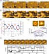
Recent advances in bioimaging with high-speed atomic force microscopy
- PMID: 32172451
- PMCID: PMC7242530
- DOI: 10.1007/s12551-020-00670-z
Recent advances in bioimaging with high-speed atomic force microscopy
Abstract
Among various microscopic techniques for characterizing protein structures and functions, high-speed atomic force microscopy (HS-AFM) is a unique technique in that it allows direct visualization of structural changes and molecular interactions of proteins without any labeling in a liquid environment. Since the development of the HS-AFM was first reported in 2001, it has been applied to analyze the dynamics of various types of proteins, including motor proteins, membrane proteins, DNA-binding proteins, amyloid proteins, and artificial proteins. This method has now become a versatile tool indispensable for biophysical research. This short review summarizes some bioimaging applications of HS-AFM reported in the last few years and novel applications of HS-AFM utilizing the unique ability of AFM to gain mechanical properties of samples in addition to structural information.
Keywords: Conformational dynamics; High-speed atomic force microscopy; Intermolecular interaction; Mechanical indentation; Single-molecule imaging.
Figures


Similar articles
-
Applications of high-speed atomic force microscopy to real-time visualization of dynamic biomolecular processes.Biochim Biophys Acta Gen Subj. 2018 Feb;1862(2):229-240. doi: 10.1016/j.bbagen.2017.07.010. Epub 2017 Jul 15. Biochim Biophys Acta Gen Subj. 2018. PMID: 28716648 Review.
-
High-speed atomic force microscopy: imaging and force spectroscopy.FEBS Lett. 2014 Oct 1;588(19):3631-8. doi: 10.1016/j.febslet.2014.06.028. Epub 2014 Jun 14. FEBS Lett. 2014. PMID: 24937145 Review.
-
High-speed atomic force microscopy and its future prospects.Biophys Rev. 2018 Apr;10(2):285-292. doi: 10.1007/s12551-017-0356-5. Epub 2017 Dec 18. Biophys Rev. 2018. PMID: 29256119 Free PMC article. Review.
-
Guide to studying intrinsically disordered proteins by high-speed atomic force microscopy.Methods. 2022 Nov;207:44-56. doi: 10.1016/j.ymeth.2022.08.008. Epub 2022 Aug 30. Methods. 2022. PMID: 36055623
-
Advances in high-speed atomic force microscopy (HS-AFM) reveal dynamics of transmembrane channels and transporters.Curr Opin Struct Biol. 2019 Aug;57:93-102. doi: 10.1016/j.sbi.2019.02.008. Epub 2019 Mar 14. Curr Opin Struct Biol. 2019. PMID: 30878714 Free PMC article. Review.
Cited by
-
Practical considerations for feature assignment in high-speed AFM of live cell membranes.Biophys Physicobiol. 2022 Apr 15;19:1-21. doi: 10.2142/biophysico.bppb-v19.0016. eCollection 2022. Biophys Physicobiol. 2022. PMID: 35797405 Free PMC article.
-
Implementation of residue-level coarse-grained models in GENESIS for large-scale molecular dynamics simulations.PLoS Comput Biol. 2022 Apr 5;18(4):e1009578. doi: 10.1371/journal.pcbi.1009578. eCollection 2022 Apr. PLoS Comput Biol. 2022. PMID: 35381009 Free PMC article.
-
Multiple dimeric structures and strand-swap dimerization of E-cadherin in solution visualized by high-speed atomic force microscopy.Proc Natl Acad Sci U S A. 2022 Jul 26;119(30):e2208067119. doi: 10.1073/pnas.2208067119. Epub 2022 Jul 22. Proc Natl Acad Sci U S A. 2022. PMID: 35867820 Free PMC article.
-
Nanotechnology meets medicine: applications of atomic force microscopy in disease.Biophys Rev. 2025 Apr 3;17(2):359-384. doi: 10.1007/s12551-025-01306-w. eCollection 2025 Apr. Biophys Rev. 2025. PMID: 40376402 Free PMC article. Review.
-
The New Era of High-Throughput Nanoelectrochemistry.Anal Chem. 2023 Jan 10;95(1):319-356. doi: 10.1021/acs.analchem.2c05105. Anal Chem. 2023. PMID: 36625121 Free PMC article. Review. No abstract available.
References
-
- Alessandrini A, Facci P. AFM: a versatile tool in biophysics. Meas Sci Technol. 2005;16:R65–R92. doi: 10.1088/0957-0233/16/6/R01. - DOI
-
- Ando T, Uchihashi T, Fukuma T. High-speed atomic force microscopy for nano-visualization of dynamic biomolecular processes. Prog Surf Sci. 2008;83:337–437. doi: 10.1016/j.progsurf.2008.09.001. - DOI
Publication types
Grants and funding
- 19H05389/Ministry of Education, Culture, Sports, Science and Technology (JP)
- 19K23737/Ministry of Education, Culture, Sports, Science and Technology (JP)
- 18H04512/Ministerio de Educación, Cultura y Deporte (ES)
- 18H01837/Ministerio de Educación, Cultura y Deporte (ES)
- 18-101/Exploratory Research Center on Life and Living Systems (JP)
LinkOut - more resources
Full Text Sources
Miscellaneous

