. 2020 Nov;40(8):1327-1338.
doi: 10.1007/s10571-020-00820-7. Epub 2020 Mar 14.

Growth-Promoting Treatment Screening for Corticospinal Neurons in Mouse and Man

Affiliations

Growth-Promoting Treatment Screening for Corticospinal Neurons in Mouse and Man

Nicholas Hanuscheck et al. Cell Mol Neurobiol. 2020 Nov.

Abstract

Neurons of the central nervous system (CNS) that project long axons into the spinal cord have a poor axon regenerative capacity compared to neurons of the peripheral nervous system. The corticospinal tract (CST) is particularly notorious for its poor regeneration. Because of this, traumatic spinal cord injury (SCI) is a devastating condition that remains as yet uncured. Based on our recent observations that direct neuronal interleukin-4 (IL-4) signaling leads to repair of axonal swellings and beneficial effects in neuroinflammation, we hypothesized that IL-4 acts directly on the CST. Here, we developed a tissue culture model for CST regeneration and found that IL-4 promoted new growth cone formation after axon transection. Most importantly, IL-4 directly increased the regenerative capacity of both murine and human CST axons, which corroborates its regenerative effects in CNS damage. Overall, these findings serve as proof-of-concept that our CST regeneration model is suitable for fast screening of new treatments for SCI.

Keywords: Corticospinal tract; Growth-promoting treatment; Interleukin-4; Regeneration; Spinal cord injury.


Conflict of interest statement

Authors declare no relevant competing interests.

Figures

Fig. 1
Fig. 1
Preparation and characterization of CxV explants. a Schematic drawing of the mouse CNS, with dorsal root ganglia (DRGs) depicted on the side of the spinal cord. Pyramidal motor neurons project their axons from primary motor cortex (M1) through the pyramidal tract into the dorsal corticospinal tract (dCST). Fibers from the DRGs ascend through the dorsal columns (DC, DRG) to the hindbrain. Sensory information is directed through several relays to the primary sensory cortex (S1). In the mouse, M1 and S1 overlap and are therefore mostly referred to as sensorimotor cortex. b Coronal 250 µm vibratome slices of a P1 mouse brain at indicated Bregma positions 0.4 to − 1.3 used for the dissection of motor cortex layer V (CxV) explants (delineated regions). Explants were divided in two or three pieces and subsequently cultivated. c Brightfield microscopic images of DRG (upper panel) and CxV (lower panel) explants show robust neurite outgrowth and stable growth cone formation (right panels) after 3 days in vitro. Axons and growth cones of the cortex explants were smaller in diameter compared to those of the DRGs. d Quantification of axon diameter of DRG and CxV axons in culture (n = 10). e Parasagittal spinal cord section of a YFP-H mouse with YFP-labeled axons of the DC and the CST. f Quantification of axon diameter of DC and CST axons in vivo (n = 10). Statistics: t-test, ***p < 0.001. Scale bars: (c) 100 µm, (e) 25 µm
Fig. 2
Fig. 2
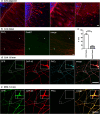
CxV explants mainly grow CST projection axons. a Immunocytochemistry of motor cortex layer V (CxV) explant cultures after 5 days in vitro for markers (red) of neurons (β-III-Tubulin, Tubb3), astrocytes (GFAP), oligodendrocytes (O4), and microglia (Iba1). Nuclei were stained with dapi (blue) and the edge of the explant delineated with white dots. b Distal CxV axons stained for Gad67 (green) and PKCγ (red) showing that the majority of the growing axons arise from CST neurons (green, arrows). c Quantification of the area of PKCy+ and Gad67+ axons relative to Tubb3 (n = 3 and images, resp.). d, e Immunocytochemistry of CxV (d) and DRG (e) explant cultures from a GFP (green) mouse at 1.6 mm distance from the explants. Axons were stained for GAP-43 (red), a marker for growing axons, and PKCγ (cyan), a marker for CST axons (boxed area enlarged). Scale bars: 25 µm. Statistics: unpaired t-test, p < 0.001
Fig. 3
Fig. 3
IL-4 increases regeneration of CST axons after transection. a Immunocytochemistry of CST axons for Tubb3 (green) and pGAP-43 (red) at 30 min after treatment with IL-4 (left panels) or PBS (right panels). b Quantification of IL-4-induced GAP-43 phosphorylation (area of pGAP-43+ pixels relative to Tubb3) (PBS n = 8; IL-4 n = 6). c CxV growth assay showing quantification induced axon outgrowth after treatment with IL-4 (n = 5) and IGF-1 (n = 4) compared to PBS controls (n = 12). d In vitro transection assay enables assessment of CST regeneration after treatment. Transection site visualized by scratch on plastic surface (left). Markings highlight regenerating (arrows), retracting (arrow heads) and degenerating (cross) axons. Axons were transected at approximately 1 mm distance to the explant (see Suppl. Fig. S2) and imaged at 0, 90, and 180 min post-transection. e Quantification of degeneration, retraction and regeneration after treatment with IGF-1 (gray bars, n = 5) and IL-4 (white bars, n = 4), compared to PBS controls (black bars, n = 5). f Immunocytochemistry of transected CST axons 120 min post-transection with Phalloidin (green) and GAP-43 (red) showing robust formation of new growth cones and thus regeneration in response to transection. Dashed line represents transection site. Scale bars: a, f 10 µm, d 20 µm. Statistics: b unpaired t-test; c, e one-way ANOVA with Tukey’s multiple comparison test *p < 0.05, ***p < 0.001
Fig. 4
Fig. 4
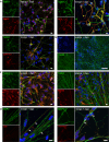
Human neurons in culture display CST markers. a Immunocytochemistry of human stem cell-derived neurons for neuronal lineage markers (NeuN, NF-L, GAP-43; all red) co-labeled with β-III-Tubulin (Tubb3, green). Nuclei labeled with dapi (blue) b Neuronal network formation after 3 weeks of differentiation. c Human neurons stained for CST marker PKCγ (red) and pyramidal neuron marker SMI-32 (red), co-labeled with Tubb3, respectively. d Synaptic contacts as shown by staining for synaptophysin (Syp, red) and Homer1 (red), with counterstaining for microtubule-associated protein 2 (MAP2, green). Scale bars: a, c, d 10 µm; b 100 µm
Fig. 5
Fig. 5
IL-4 induces neurite outgrowth of human neurons. a Human neurons express the IL-4Rα (red) as shown by overlap with the neuronal marker β-III-Tubulin (Tubb3) (green). b Exemplary images of IRS1 and pIRS stainings in PBS and IL-4 treated cultures. c Quantification of fluorescence intensity ratio of pIRS1 and IRS1 in Tubb3+ cells (n = 10 images per treatment). d Exemplary images of human neurons treated with IL-4, IL-4 + αIL-4 antibody, or PBS for 24 h stained with Tubb3 (cyan) and dapi (blue). e Quantification of treatment effects for 24 h shows a significant increase in Tubb3+ neurite outgrowth in IL-4-treated cultures that is abolished by αIL-4 (n = 100–150 neurons per treatment). Scale bars: a, b 10 µm, d 50 µm. Statistics: c unpaired t-test and e one-way ANOVA with Tukey’s multiple comparison test ***p < 0.001

Similar articles

Cited by

  • Interleukin-4 receptor signaling modulates neuronal network activity.
    Hanuscheck N, Thalman C, Domingues M, Schmaul S, Muthuraman M, Hetsch F, Ecker M, Endle H, Oshaghi M, Martino G, Kuhlmann T, Bozek K, van Beers T, Bittner S, von Engelhardt J, Vogt J, Vogelaar CF, Zipp F. Hanuscheck N, et al. J Exp Med. 2022 Jun 6;219(6):e20211887. doi: 10.1084/jem.20211887. Epub 2022 May 19. J Exp Med. 2022. PMID: 35587822 Free PMC article.

References

    1. Abu-Rub M, McMahon S, Zeugolis DI, Windebank A, Pandit A (2010) Spinal cord injury in vitro: modelling axon growth inhibition. Drug Discov Today 15:436–443. 10.1016/j.drudis.2010.03.008 - PubMed
    1. Bagnard D, Chounlamountri N, Puschel AW, Bolz J (2001) Axonal surface molecules act in combination with semaphorin 3a during the establishment of corticothalamic projections. Cereb Cortex 11:278–285 - PubMed
    1. Bradke F, Fawcett JW, Spira ME (2012) Assembly of a new growth cone after axotomy: the precursor to axon regeneration. Nat Rev Neurosci 13:183–193. 10.1038/nrn3176 - PubMed
    1. Bunge MB (2008) Novel combination strategies to repair the injured mammalian spinal cord. J Spinal Cord Med 31:262–269 - PMC - PubMed
    1. Carter LM, Starkey ML, Akrimi SF, Davies M, McMahon SB, Bradbury EJ (2008) The yellow fluorescent protein (YFP-H) mouse reveals neuroprotection as a novel mechanism underlying chondroitinase ABC-mediated repair after spinal cord injury. J Neurosci 28:14107–14120. 10.1523/jneurosci.2217-08.2008 - PMC - PubMed

MeSH terms

Substances