. 2020 May;75(5):1305-1314.
doi: 10.1161/HYPERTENSIONAHA.119.14608. Epub 2020 Mar 16.

Effects of Chronic Nicotine Inhalation on Systemic and Pulmonary Blood Pressure and Right Ventricular Remodeling in Mice

Affiliations

Effects of Chronic Nicotine Inhalation on Systemic and Pulmonary Blood Pressure and Right Ventricular Remodeling in Mice

Joshua M Oakes et al. Hypertension. 2020 May.

Abstract

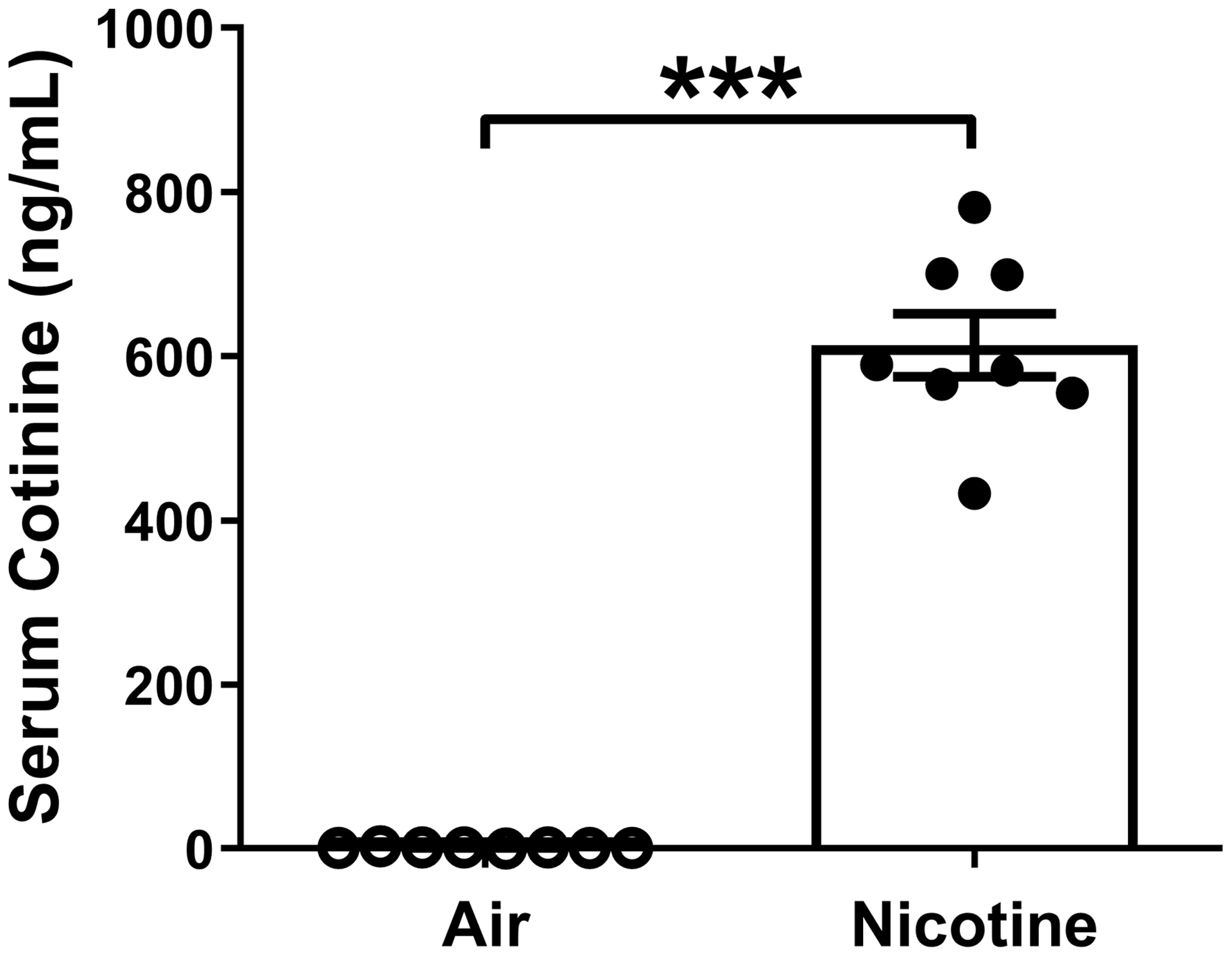
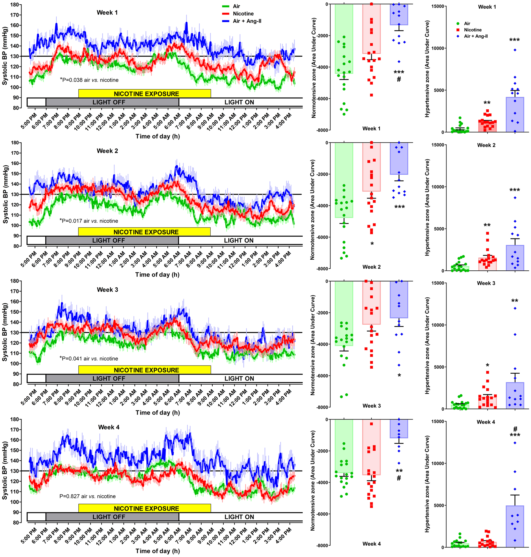
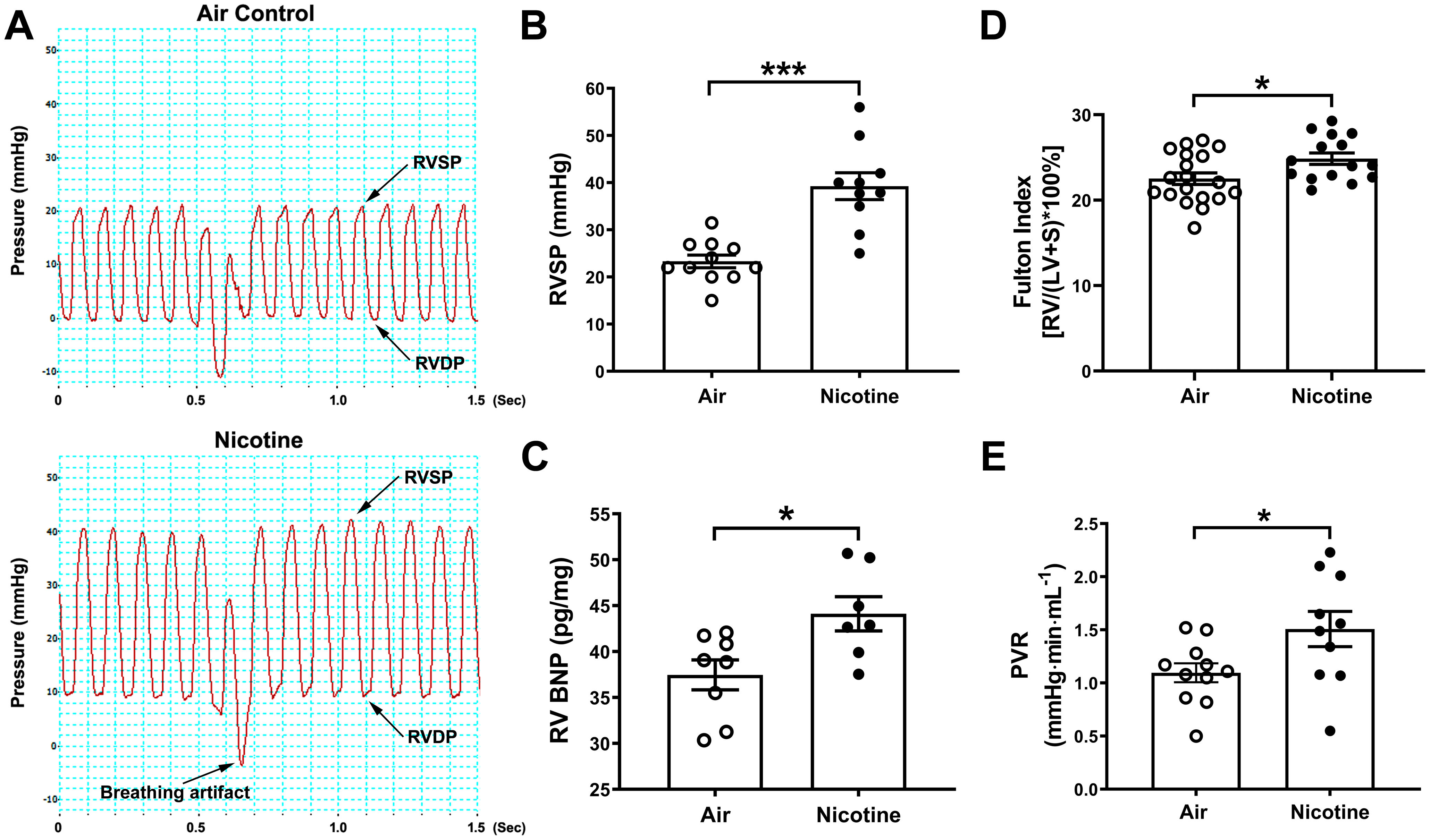
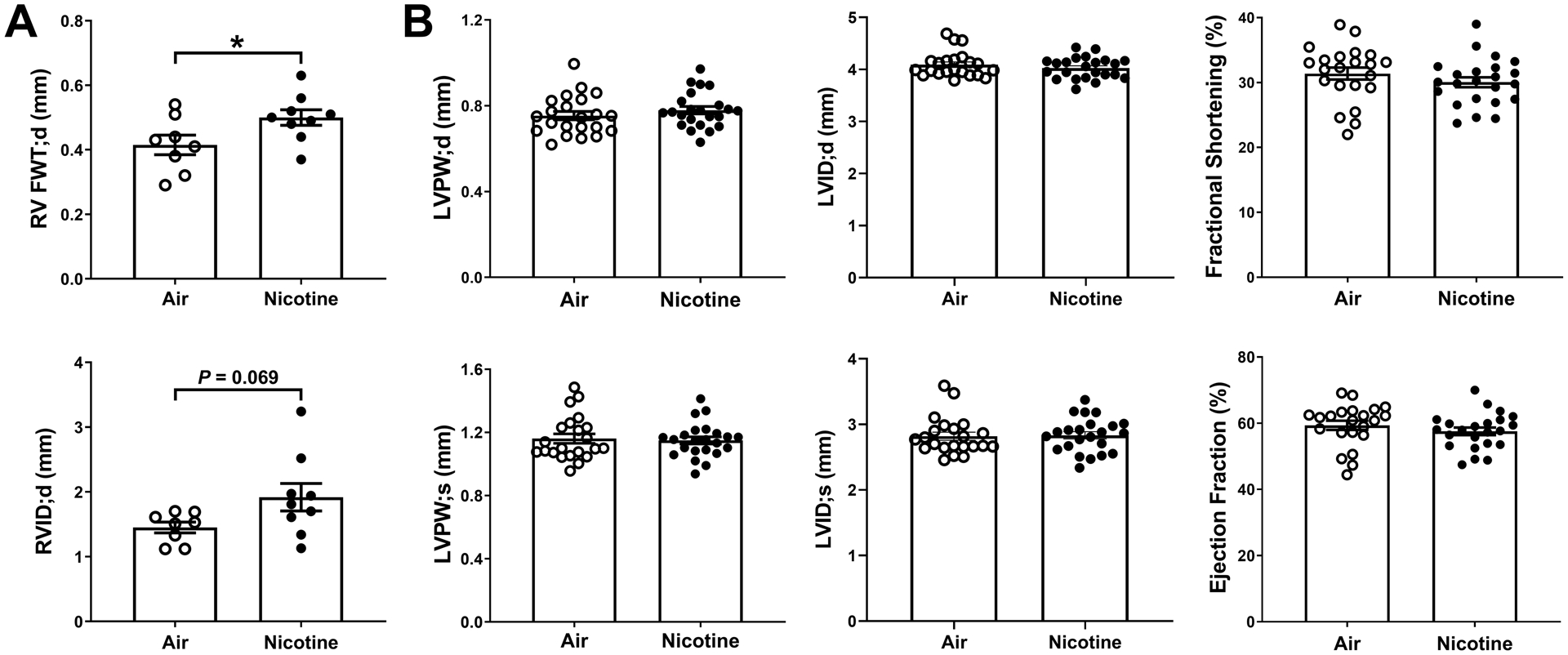
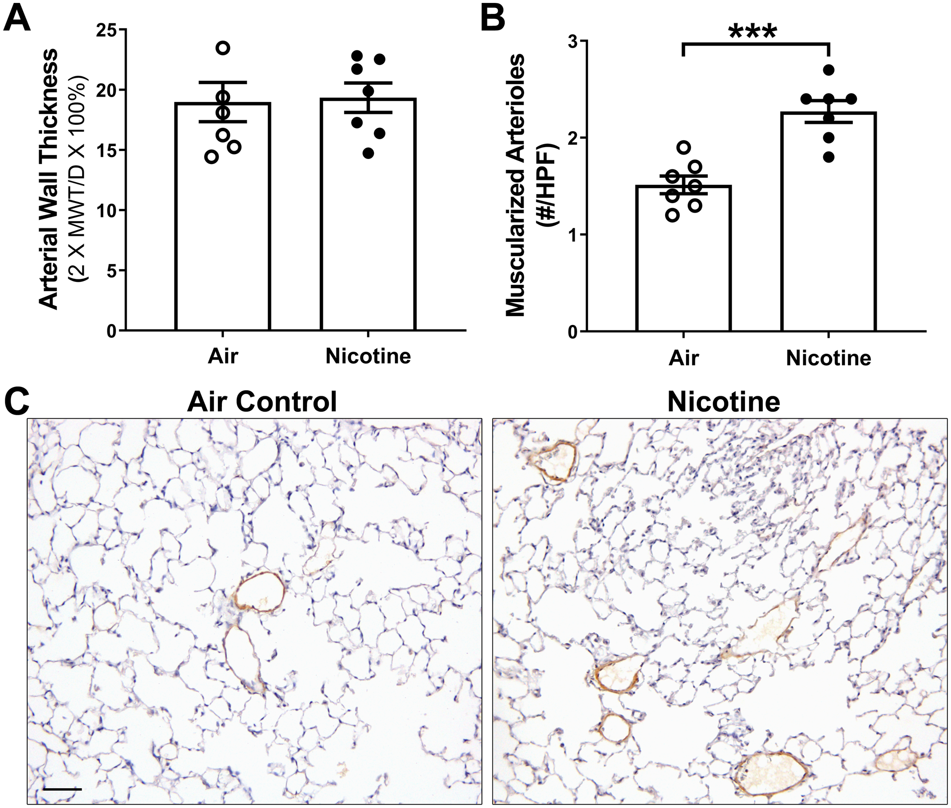
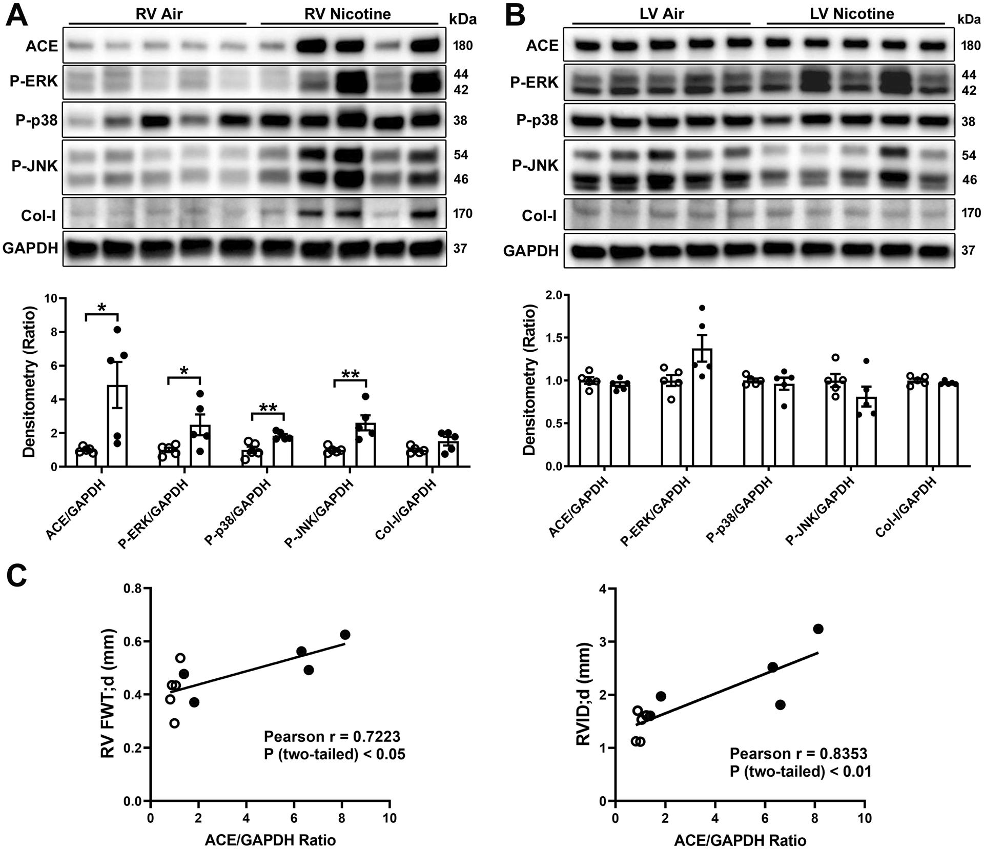
Cigarette smoking is the single most important risk factor for the development of cardiovascular and pulmonary diseases; however, the role of nicotine in the pathogenesis of these diseases is incompletely understood. The purpose of this study was to examine the effects of chronic nicotine inhalation on the development of cardiovascular and pulmonary disease with a focus on blood pressure and cardiac remodeling. Male C57BL6/J mice were exposed to air (control) or nicotine vapor (daily, 12 hour on/12 hour off) for 8 weeks. Systemic blood pressure was recorded weekly by radio-telemetry, and cardiac remodeling was monitored by echocardiography. At the end of the 8 weeks, mice were subjected to right heart catheterization to measure right ventricular systolic pressure. Nicotine-exposed mice exhibited elevated systemic blood pressure from weeks 1 to 3, which then returned to baseline from weeks 4 to 8, indicating development of tolerance to nicotine. At 8 weeks, significantly increased right ventricular systolic pressure was detected in nicotine-exposed mice compared with the air controls. Echocardiography showed that 8-week nicotine inhalation resulted in right ventricular (RV) hypertrophy with increased RV free wall thickness and a trend of increase in RV internal diameter. In contrast, there were no significant structural or functional changes in the left ventricle following nicotine exposure. Mechanistically, we observed increased expression of angiotensin-converting enzyme and enhanced activation of mitogen-activated protein kinase pathways in the RV but not in the left ventricle. We conclude that chronic nicotine inhalation alters both systemic and pulmonary blood pressure with the latter accompanied by RV remodeling, possibly leading to progressive and persistent pulmonary hypertension.

Keywords: angiotensin-converting enzyme; blood pressure; hypertension; hypertension, pulmonary; nicotine.


Figures

Figure 1.
Figure 1.
Serum cotinine levels in air- and nicotine-exposed mice. Weekly averages from air- or nicotine-exposed mice (5–6/group) during the 8-week exposure period (n=8) were shown. ***P < 0.001, nicotine vs. air control group (unpaired student t-test).
Figure 2.
Figure 2.
Chronic nicotine inhalation leads to early increase in systolic blood pressure (BP). Weekly average 24 h recording of systolic BP in mice exposed to air (green, n=18) or nicotine (red, n=18). Some mice were infused with angiotensin-II (450 ng/kg/min) as a hypertensive positive control (blue, n=9–12). Results of the two-way ANOVA between air-and nicotine-exposed mice are indicated below the traces. Area under the curve was calculated for the BP traces below and above the 130 mmHg threshold to identify normotensive and hypertensive zones, respectively, as defined in the Methods section. Statistical significance: *P<0.05, **P<0.01, ***P<0.001 vs. air control group and #P<0.05 vs. nicotine-exposed mice (Kruskal-Wallis test followed by Dunn’s multiple comparison test).
Figure 3.
Figure 3.
Chronic nicotine inhalation leads to the development of pulmonary hypertension. (A) Representative right ventricular (RV) pressure tracing from air- (upper) and nicotine- (lower) exposed mice. RVSP, RV systolic pressure; RVDP, RV diastolic pressure. (B) Quantification of RVSP. (C) Expression of brain or B-type natriuretic peptide (BNP) in RV protein extracts measured by ELISA. (D) Fulton Index. S, interventricular septum. (E) Pulmonary vascular resistance (PVR). *P<0.05, ***P<0.001, nicotine vs. air control group (unpaired student t-test).
Figure 4.
Figure 4.
Chronic nicotine inhalation leads to RV, but not LV, remodeling as revealed by echocardiography. (A) RV measurements, including RV free wall thickness (FWT) and RV internal diameter (RVID) at diastole (d). Air, n=8; Nicotine, n=9. *P<0.05, nicotine vs. air control group (unpaired student t-test). (B) LV measurements, including LV posterior wall (LVPW) and LVID at both diastole (d) and systole (s), fractional shortening and ejection fraction. Air, n=22; Nicotine, n=23.
Figure 5.
Figure 5.
Chronic nicotine inhalation promotes pulmonary vascular remodeling. (A) Arterial wall thickness was measured as 2 × medial wall thickness (MWT)/external diameter (D) × 100%. Measurements were performed on lung tissue sections immunostained with α-SMA and all arterioles associated with the bronchioles with internal diameters between 30 to 100 μm were included. Air, n=7; nicotine, n=7. (B) Number of α-SMA positive, muscularized arterioles in the alveolar region in air- and nicotine-exposed mice per high power field (HPF). The average of 10 HPFs from each mouse was used. Air, n=7; nicotine, n=7. ***P<0.001, nicotine vs. air control group (unpaired student t-test). (C) Representative α-SMA immunostaining (brown) of lung sections from air- and nicotine-exposed mice. Scale bar, 50 μm.
Figure 6.
Figure 6.
Chronic nicotine inhalation leads to ACE overexpression and MAPK activation in the RV, but not in the LV. (A) Representative Western blots of RV protein extracts and Western densitometry quantification. (B) Representative Western blots of LV protein extracts and Western densitometry quantification. Col-I, collagen I. Open circles, air; closed black circles, nicotine. *P<0.05, **P<0.01, nicotine vs. air control group (unpaired student t-test). Protein expression levels were normalized to the expression of GAPDH. For phosphorylated MAPKs, normalization to respective total MAPKs gave similar results (not shown). (C) Correlation between RV ACE expression and RV free wall thickness (FWT) and RV internal diameter (RVID) at diastole (d).

Similar articles

Cited by

References

    1. Jamal A, Phillips E, Gentzke AS, Homa DM, Babb SD, King BA, Neff LJ. Current cigarette smoking among adults - united states, 2016. MMWR Morb Mortal Wkly Rep. 2018;67:53–59 - PMC - PubMed
    1. Wagoner KG, Cornacchione J, Wiseman KD, Teal R, Moracco KE, Sutfin EL. E-cigarettes, hookah pens and vapes: Adolescent and young adult perceptions of electronic nicotine delivery systems. Nicotine Tob Res. 2016;18:2006–2012 - PMC - PubMed
    1. García Calzado MC, García Rojas JF, Mangas Rojas A, Millán J. Tobacco and arterial pressure (ii.). The acute effects on the angiotensin-converting enzyme. An Med Interna. 1990;7:392–395 - PubMed
    1. Groppelli A, Giorgi DM, Omboni S, Parati G, Mancia G. Persistent blood pressure increase induced by heavy smoking. Journal of hypertension. 1992;10:495–499 - PubMed
    1. Cellina GU, Honour AJ, Littler WA. Direct arterial pressure, heart rate, and electrocardiogram during cigarette smoking in unrestricted patients. Am Heart J. 1975;89:18–25 - PubMed

Publication types

MeSH terms