

Case Reports
doi: 10.1080/13816810.2020.1737949.
Epub 2020 Mar 16.
Quasidominance in autosomal recessive RDH12-Leber congenital amaurosis
Affiliations
- PMID: 32172635
- PMCID: PMC7513925
- DOI: 10.1080/13816810.2020.1737949
Item in Clipboard
Case Reports
Quasidominance in autosomal recessive RDH12-Leber congenital amaurosis
Ophthalmic Genet.
2020 Apr.
No abstract available
Figures

A 34-year old man (Patient 1, IV-2) visited our clinic with his 3-year old daughter (Patient 2, V-3). Clinical presentation of both patients was suggestive of a diagnosis of Leber Congenital Amaurosis caused by the gene RDH12. Family history was significant for multiple consanguineous marriages.
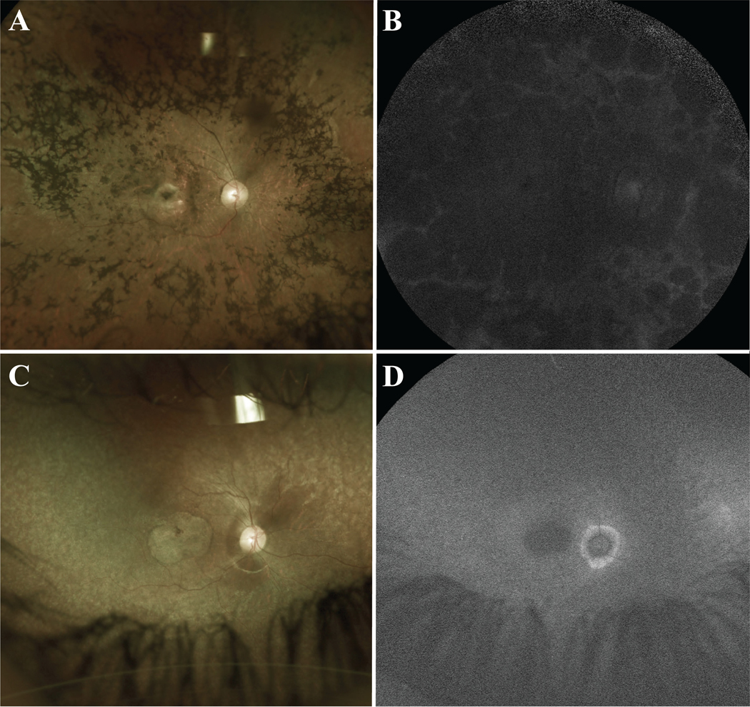
Color photography of the posterior pole of Patient 1 (A and B) revealed dense and widespread intraretinal pigment migration, a severely atrophic and demarcated macula with pigmentation and yellowing, optic disc pallor, attenuation of the retinal vessels, and generalized retinal atrophy (A). On short-wave fundus autofluorescence (SW-AF) images, generalized hypoautofluorescence was observed throughout the posterior pole, corresponding to atrophic retinal pigment epithelium (B). In Patient 2 (C and D) color photography revealed a severely atrophic and demarcated macula, along with mottling of the retinal pigment epithelium (RPE) throughout the posterior pole, optic disc pallor, and attenuation of the retinal vessels. Generalized retinal atrophy with preservation of the peripapillary area was also appreciated (C). SW-AF images also revealed a generalized loss of autofluorescence. The atrophic macula appeared as an area of dense hypoautofluorescence, with peripapillary sparing of the RPE evident as relative higher levels of autofluorescence surrounding the optic disc as compared to the rest of the fundus with generalized hypoautofluorescence (D).
Similar articles
-
Novel RDH12 sequence variations in Leber congenital amaurosis.J AAPOS. 2010 Aug;14(4):349-51. doi: 10.1016/j.jaapos.2010.04.010. J AAPOS. 2010. PMID: 20736127
-
Peripapillary sparing in RDH12-associated Leber congenital amaurosis.Ophthalmic Genet. 2017 Dec;38(6):575-579. doi: 10.1080/13816810.2017.1323339. Epub 2017 May 17. Ophthalmic Genet. 2017. PMID: 28513254 Free PMC article.
-
Genome-wide homozygosity mapping in families with leber congenital amaurosis identifies mutations in AIPL1 and RDH12 genes.DNA Cell Biol. 2014 Dec;33(12):876-83. doi: 10.1089/dna.2014.2554. DNA Cell Biol. 2014. PMID: 25148430
-
Retinol dehydrogenase 12 (RDH12): Role in vision, retinal disease and future perspectives.Exp Eye Res. 2019 Nov;188:107793. doi: 10.1016/j.exer.2019.107793. Epub 2019 Sep 7. Exp Eye Res. 2019. PMID: 31505163 Review.
-
A Mini-review: Animal Models of GUCY2D Leber Congenital Amaurosis (LCA1).Adv Exp Med Biol. 2016;854:253-8. doi: 10.1007/978-3-319-17121-0_34. Adv Exp Med Biol. 2016. PMID: 26427419 Review.
Cited by
-
Primary versus Secondary Elevations in Fundus Autofluorescence.Int J Mol Sci. 2023 Aug 2;24(15):12327. doi: 10.3390/ijms241512327. Int J Mol Sci. 2023. PMID: 37569703 Free PMC article. Review.
-
Bisretinoid lipofuscin, fundus autofluorescence and retinal disease.Prog Retin Eye Res. 2025 Jul 8;108:101388. doi: 10.1016/j.preteyeres.2025.101388. Online ahead of print. Prog Retin Eye Res. 2025. PMID: 40639497 Review.
-
Iron overload and chelation modulates bisretinoid levels in the retina.Front Ophthalmol (Lausanne). 2023 Dec 22;3:1305864. doi: 10.3389/fopht.2023.1305864. eCollection 2023. Front Ophthalmol (Lausanne). 2023. PMID: 38983013 Free PMC article.
References
-
- Jauregui R, Cho GY, Takahashi VKL, Takiuti JT, Bassuk AG, Mahajan VB, et al. Caring for Hereditary Childhood Retinal Blindness. Asia Pac J Ophthalmol (Phila). 2018. - PubMed
-
- den Hollander AI, Roepman R, Koenekoop RK, Cremers FP. Leber congenital amaurosis: genes, proteins and disease mechanisms. Prog Retin Eye Res. 2008;27(4):391–419. - PubMed
-
- Aleman TS, Uyhazi KE, Serrano LW, Vasireddy V, Bowman SJ, Ammar MJ, et al. RDH12 Mutations Cause a Severe Retinal Degeneration With Relatively Spared Rod Function. Invest Ophthalmol Vis Sci. 2018;59(12):5225–36. - PubMed
Publication types
MeSH terms
Substances
Grants and funding
LinkOut - more resources
Full Text Sources