Review
. 2020;18(8):674-695.
doi: 10.2174/1570159X18666200203101828.

A Comprehensive Review of Alzheimer's Association with Related Proteins: Pathological Role and Therapeutic Significance

Affiliations
Review

A Comprehensive Review of Alzheimer's Association with Related Proteins: Pathological Role and Therapeutic Significance

Deepak Kumar et al. Curr Neuropharmacol. 2020.

Abstract

Alzheimer's is an insidious, progressive, chronic neurodegenerative disease which causes the devastation of neurons. Alzheimer's possesses complex pathologies of heterogeneous nature counting proteins as one major factor along with enzymes and mutated genes. Proteins such as amyloid precursor protein (APP), apolipoprotein E (ApoE), presenilin, mortalin, calbindin-D28K, creactive protein, heat shock proteins (HSPs), and prion protein are some of the chief elements in the foremost hypotheses of AD like amyloid-beta (Aβ) cascade hypothesis, tau hypothesis, cholinergic neuron damage, etc. Disturbed expression of these proteins results in synaptic dysfunction, cognitive impairment, memory loss, and neuronal degradation. On the therapeutic ground, attempts of developing anti-amyloid, anti-inflammatory, anti-tau therapies are on peak, having APP and tau as putative targets. Some proteins, e.g., HSPs, which ameliorate oxidative stress, calpains, which help in regulating synaptic plasticity, and calmodulin-like skin protein (CLSP) with its neuroprotective role are few promising future targets for developing anti-AD therapies. On diagnostic grounds of AD C-reactive protein, pentraxins, collapsin response mediator protein-2, and growth-associated protein-43 represent the future of new possible biomarkers for diagnosing AD. The last few decades were concentrated over identifying and studying protein targets of AD. Here, we reviewed the physiological/pathological roles and therapeutic significance of nearly all the proteins associated with AD that addresses putative as well as probable targets for developing effective anti-AD therapies.

Keywords: Alzheimer`s; neurodegeneration; pathological role; proteins; therapeutics.


Figures

Fig. (1)
Fig. (1)
Locations of the proteins in major brain areas (Cerebral cortex, Hippocampus, Cerebellum, Caudate). (A higher resolution / colour version of this figure is available in the electronic copy of the article).
Fig. (2)
Fig. (2)
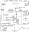
Schematic representation of proteins associated with neurotoxicity and neurodegeneration. AD is a disease caused by a complex talk between various pathologies and proteins associated with them. For example, amyloidogenic processing of APP results in the secretion of one major form of Aβ, i.e., Aβ-40, along with few minor species containing 42 or 43 amino acid residues, which later on gets aggregated in the brain . Increased Aβ accumulation in the brain enhances APP/Go protein complex formation, which then initiates Go-GβΥ complex signaling and as a result, irregular activation of the MPAK-p38 pathway causes neurotoxicity. MCP-1 is primarily expressed by glial cells, i.e., astrocytes and microglia. Astrocytes rapidly express MCP-1 in ipsilateral thalamus as a primary response to brain injury. Later on, MCP-1 acts as a potent chemoattractant for monocytes, memory T cells, and dendritic cells, as a result, it initiates a cascade of secondary responses in inflammation and causes neurodegeneration through direct apoptosis. ILs are also secreted during various pathological processes and further enhance neuroinflammation, e.g., IL-1 activates T cells and other inflammatory mediators, which initiate an inflammatory response and activate p38-MAPK. With the initiation of inflammation, neutrophils and macrophages secrete IL-6, which in turn, facilitates the production of C-reactive protein. C-reactive protein further promotes inflammation and increases the production of Aβ-42. Despite this, ubiquilin-1 facilitates the accumulation of insoluble Aβ peptides by affecting BACE1 activity and via assisting presenilin 1 accumulation, which afterward, results in presenilin-dependent γ-secretase degradation of APP. Copper-related protein CutA also interferes with BACE1 activity and indirectly affects APP processing. In AD, iron levels have been found to be elevated and facilitating cognitive decline. Mitoferrin-1 is associated with iron homeostasis and its up-regulation contributes to the excessive accumulation of iron in mitochondria, resulting in increased mitochondria induced oxidative stress. Moreover, aggregation of alpha-synuclein as Lewy bodies is potentiated by ganglioside lipids, which results in increased alpha-synuclein-mediated oxidative stress, neurotoxicity, and ultimately neurodegeneration. From CNS, Aβ is cleared out via perivascular circulation and glymphatic pathway where P-glycoprotein and LRP-1 along with SORLA facilitate its transportation. Disrupted functioning of SORLA prevents Aβ clearance from the brain. Moreover, SORLA facilitates calpain mediated proteolytic degradation of synapsins and thus, potentiating impaired regulation of neurotransmitters release at synapsis. In addition, overactivation of calpain-2 puts restrictions to the extent of LTP and aids Aβ accumulation via supplementing the expression level of β-secretase. On the other hand, CIP2A and CDK5RAP2 facilitate hyperphosphorylation of tau protein; as a result, disruption of nucleocytoplasmic transport causes neuronal death and NFTs. In addition, ApoE4’s isoform-specific interactions with tau, PrPC-induced neurotoxicity, and disrupted Gap43 activities (such as synaptic transmission, membrane permeability, neuronal growth, etc.) also cause an upsurge in NFTs level in the brain. However, the role of CSPGs is remained unexplored yet; they are also found abundantly in Aβ-plaques and NFTs. (A higher resolution / colour version of this figure is available in the electronic copy of the article).
Fig. (3)
Fig. (3)
Role of presenilin in neurodegeneration. Presenilin proteins, i.e., presenilin 1 and presenilin 2 (along with other proteins), are structural units of γ-secretase complex. In the γ-secretase protein complex, presenilin acts as an active site after being cleaved into an N- and C-terminal fragment. Moreover, it helps the γ-secretase complex to exert its activity by letting the substrate pass through the hydrophilic pocket in the membrane formed by two presenilin fragments. Within the hydrophobic environment of the plasma membrane, γ-secretase cleaves the C-terminal fragment of APP after it gets cleaved by α- or β-secretase (in non-amyloidogenic and amyloidogenic pathways, respectively) to release the APP’s cytoplasmic domain. As a key factor in γ-secretase activity, the mutation in presenilin can facilitate overactivation of the amyloidogenic pathway and as a result, the production of Aβ–42 gets enhanced in the brain. In addition, ubiquilin-1 fuels this pathology more aggressively as it facilitates the presenilin 1 accumulation and aggresome formation by ubiquilin-1 transcript variant 1 and ubiquilin-1 transcript variant 2. (A higher resolution / colour version of this figure is available in the electronic copy of the article).
Fig. (4)
Fig. (4)
Schematic representation of proteins associated with neuroprotection and regeneration. These proteins help in the growth, regulation, and protection of neurons. CRMP-2, after getting phosphorylated by Rho-kinase and glycogen synthase, assists axon development, and binds/regulate microtubules assembly, respectively. Along with CRMP-2, GAP-43 also contributes to the growth of axons and modulates the formation of new connections. However, because of high lipid content and oxygen consumption in the brain, there is an increased amount of oxidative stress. In order to counteract this situation, brain expresses HSPs in different regions of CNS such as cortical region (especially in astrocytes). These HSPs also ameliorate the decline in Purkinje cells of the brain and halt neurodegeneration. CLSP exhibits neuroprotection by interacting with htHNR and inhibiting the loss of synaptophysin, which indeed facilitate detoxification and synaptic transmission via playing a crucial role in the biogenesis of secretory vesicles and prompting the targeting of VMATs towards these vesicles. In addition, calpain-1 also plays an important role in the initiation of LTP. On the other hand, ubiquitin helps in restoring the γ-secretase activity, clearing denatured protein fragments and insoluble aggregates such as Aβ plaques and NFTs via ubiquitin-dependent protein degradation system. P-glycoprotein and LRP-1, along with SORLA, also assist Aβ clearance and facilitate its transportation across the BBB. However, Aβ mediated microglia activation and therefore the expression of TNF-α, IL-1β, and NF-κB cause reduction in the expression of P-glycoprotein, which later is halted by Mortalin overexpression, as it extenuates Aβ mediated cell damage and apoptosis via inhibiting the mPTP, caspase-3 activation, and release of pro-apoptotic factor cytochrome C. Moreover, overexpressed mortalin, along with calbindin-D28k, regulates intracellular calcium concentrations, which help in reducing the oxidative stress and glutamate neurotoxicity. Moreover, increased RANTES expression is associated with the activation of PI-3K and MAPK signaling pathways and this phenomenon exhibits neuroprotection. However, the role of neurogranin in neuroprotection is yet to be discovered, it is believed to be facilitating the hippocampal synaptic plasticity and spatial learning. (A higher resolution / colour version of this figure is available in the electronic copy of the article).
Fig. (5)
Fig. (5)
Role of insulin in neuroprotection. Insulin helps in halting Aβ degradation by increasing neprilysin secretion and expression of insulin-degrading enzyme in astrocytes via the activation of the ERK-mediated pathway. However, impaired Biliverdin reductase-A, increased mTOR activity, and Aβ accumulation in the brain result in impaired insulin regulation and downregulation of plasma membrane insulin receptors, respectively. Insulin resistance and impaired insulin regulation also result in overactivation of GSK-3β, which ultimately takes part in the hyperphosphorylation of tau protein and generation of NFTs. (A higher resolution / colour version of this figure is available in the electronic copy of the article).

Similar articles

Cited by

References

    1. Scheltens P., Blennow K., Breteler M.M., de Strooper B., Frisoni G.B., Salloway S., Van der Flier W.M. Alzheimer’s disease. Lancet. 2016;388(10043):505–517. doi: 10.1016/S0140-6736(15)01124-1. - DOI - PubMed
    1. World Health Organization and Alzheimer’s Disease International Dementia: a public health priority. 2012.
    1. WHO Dementia Fact sheet. 2017 https://www.who.int/en/news-room/fact-sheets/detail/dementia
    1. Alzheimer’s & Related Disorders Society of India . The Dementia India Report: prevalence, impact, costs and services for Dementia: Executive Summary. New Delhi: ARDSI; 2010.
    1. Alzheimer’s Association 2018 Alzheimer’s disease Facts and Figures. Alzheimers Dement. 2018;14:367–429. doi: 10.1016/j.jalz.2018.02.001. - DOI