

DNA damage signaling in the cellular responses to mustard vesicants
- PMID: 32173488
- PMCID: PMC7224631
- DOI: 10.1016/j.toxlet.2020.03.008
DNA damage signaling in the cellular responses to mustard vesicants
Abstract
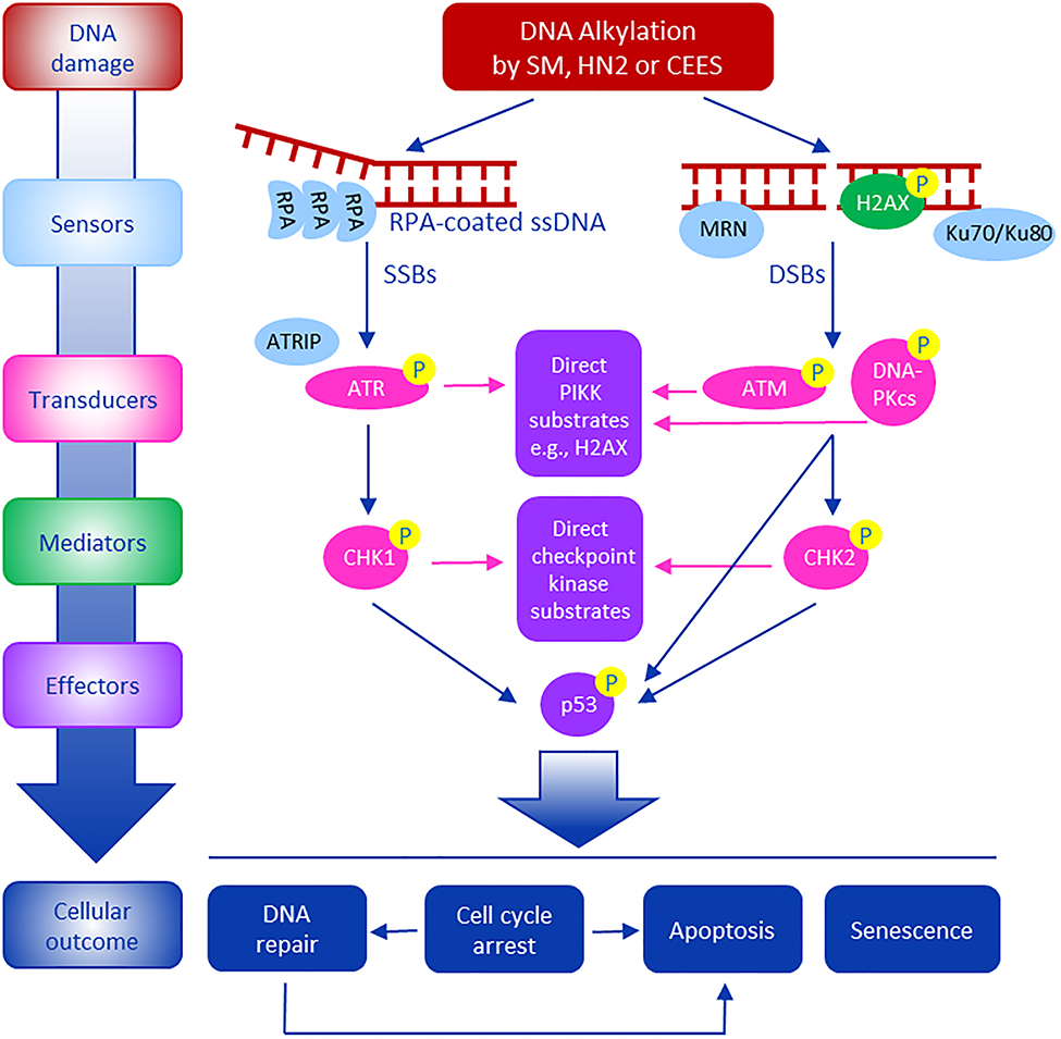
Mustard vesicants, including sulfur mustard (2,2'-dichlorodiethyl sulfide, SM) and nitrogen mustard (bis(2-chloroethyl)methylamine, HN2) are cytotoxic blistering agents synthesized for chemical warfare. Because they contain highly reactive electrophilic chloroethyl side chains, they readily react with cellular macromolecules like DNA forming monofunctional and bifunctional adducts. By targeting DNA, mustards can compromise genomic integrity, disrupt the cell cycle, and cause mutations and cytotoxicity. To protect against genotoxicity following exposure to mustards, cells initiate a DNA damage response (DDR). This involves activation of signaling cascades including ATM (ataxia telangiectasia mutated), ATR (ataxia telangiectasia and Rad3-related) and DNA-PKcs (DNA-dependent protein kinase, catalytic unit). Signaling induced by the DDR leads to the recruitment and activation of repair related proteins such as phospho H2AX and phospho p53 to sites of DNA lesions. Excessive DNA modifications by mustards can overwhelm DNA repair leading to single and double strand DNA breaks, cytotoxicity and tissue damage, sometimes leading to cancer. Herein we summarize DDR signaling pathways induced by SM, HN2 and the half mustard, 2-chloroethyl ethyl sulfide (CEES). At the present time, little is known about how mustard-induced DNA damage leads to the activation of DDR signaling. A better understanding of mechanisms by which mustard vesicants induce the DDR may lead to the development of countermeasures effective in mitigating tissue injury.
Keywords: Cell cycle; DNA damage response; H2AX; Nitrogen mustard; Sulfur mustard; Vesicants; p53.
Copyright © 2020 Elsevier B.V. All rights reserved.
Conflict of interest statement
Declaration of Competing Interest
The authors declare that they have no known competing financial interests or personal relationships that could have appeared to influence the work reported in this paper.
Figures
References
-
- Balcome S, Park S, Quirk Dorr DR, Hafner L, Phillips L, Tretyakova N, 2004. Adenine-containing DNA-DNA cross-links of antitumor nitrogen mustards. Chem. Res. Toxicol 17, 950–962. - PubMed
-
- Bhat KR, Benton BJ, Ray R, 2006. Poly (ADP-ribose) polymerase (PARP) is essential for sulfur mustard-induced DNA damage repair, but has no role in DNA ligase activation. J. Appl. Toxicol 26, 452–457. - PubMed
-
- Black AT, Hayden PJ, Casillas RP, Heck DE, Gerecke DR, Sinko PJ, Laskin DL, Laskin JD, 2010. Expression of proliferative and inflammatory markers in a full-thickness human skin equivalent following exposure to the model sulfur mustard vesicant, 2-chloroethyl ethyl sulfide. Toxicol. Appl. Pharmacol 249, 178–187. - PMC - PubMed
-
- Blackford AN, Jackson SP, 2017. ATM, ATR, and DNA-PK: the trinity at the heart of the DNA damage response. Mol. Cell 66, 801–817. - PubMed
MeSH terms
Substances
Grants and funding
LinkOut - more resources
Full Text Sources
Research Materials
Miscellaneous
