. 2020 May 22;61(3):388-398.
doi: 10.1093/jrr/rraa009.

Protective effects of new aryl sulfone derivatives against radiation-induced hematopoietic injury

Affiliations

Protective effects of new aryl sulfone derivatives against radiation-induced hematopoietic injury

Jian Cao et al. J Radiat Res. .

Abstract

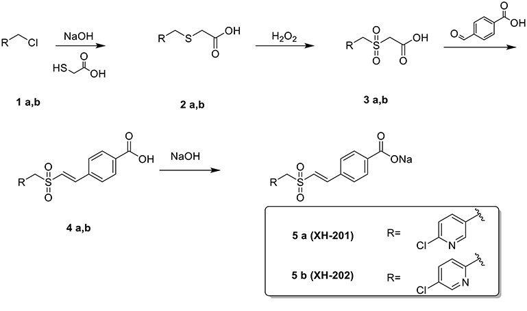
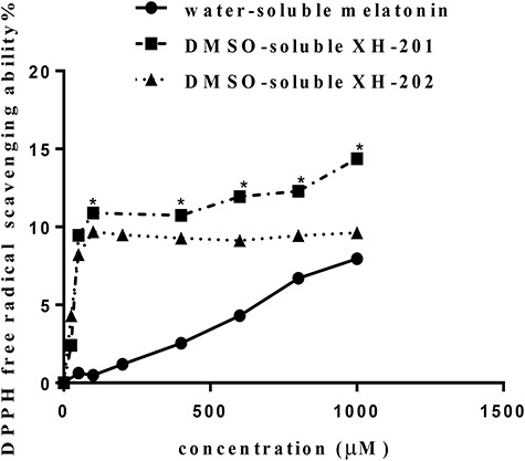
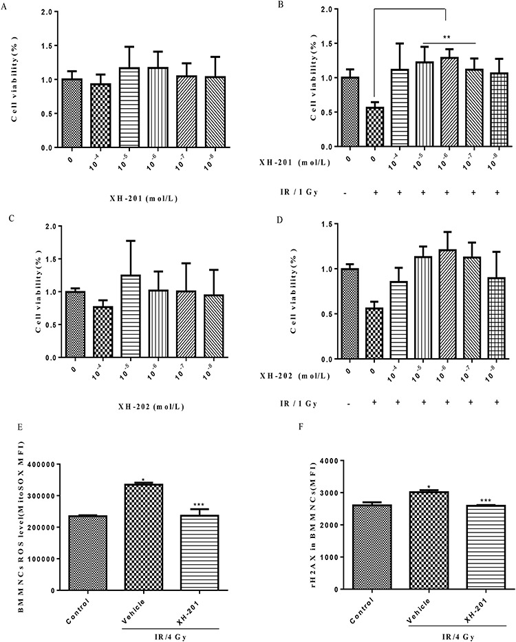
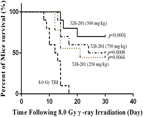
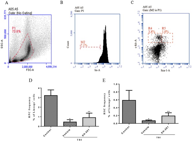
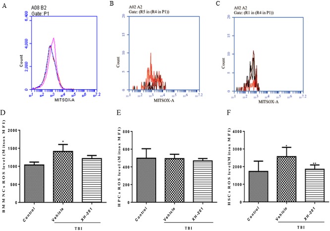
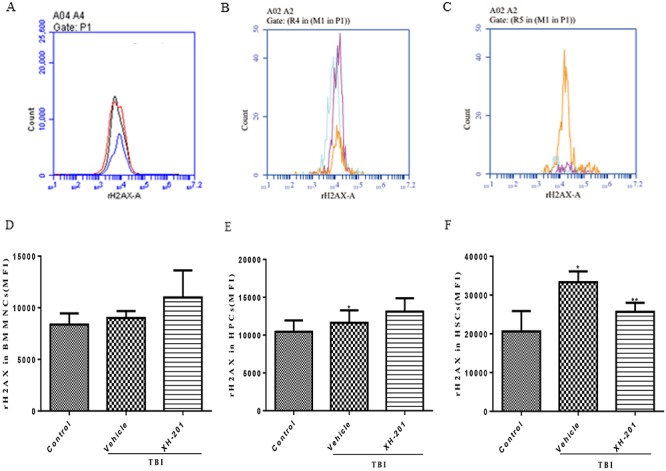
The hematopoietic system is sensitive to radiation. In this research, new aryl sulfone derivatives (XH-201 and XH-202) containing a nitrogen heterocycle were designed and synthesized and their radio-protective efficacies with regard to the hematopoietic system were evaluated. XH-201 administration significantly increased the survival rate of mice after 8.0 Gy total body irradiation (TBI). The results showed that XH-201 treatment not only increased the white blood cells, platelets counts and the percentage of hematopoietic progenitor cells and hematopoietic stem cells in mice exposed to 4.0 Gy TBI but also decreased DNA damage, as determined by flow cytometric analysis of histone H2AX phosphorylation. In addition, our data demonstrated that XH-201 decreased the mitochondrial reactive oxygen species (ROS) levels in hematopoietic cells. Overall, these data suggest that XH-201 is beneficial for the protection of the hemoatopoietic system against radiation-induced injuries.

Keywords: aryl sulfone derivatives; hematopoietic cells; radioprotection.


Figures

Scheme 1.
Scheme 1.
Preparation of XH-201 and XH-202
Fig. 1.
Fig. 1.
Effects on DPPH free radical scavenging ability of different compounds (XH-201, XH-202 and melatonin). Eight different concentrations of XH-201, XH-202 and melatonin (from 25 μM to 1000 μM) were detected by DPPH at 517 nm. *Significant difference vs melatonin (P < 0.05).
Fig. 2.
Fig. 2.
Evaluation of the radioprotective activity and cytotoxicity of XH-201 and XH-202 in vitro. The cells were sham-irradiated as a control or irradiated with 1.0 Gy after receiving different concentrations of XH-201 and cultured for 18 h. Cell viability was monitored using the luminescence assay, as described in the text. The other part cells were sham-irradiated as a control or irradiated with 4.0 Gy after receiving 10−6 mol/L XH-201 and after 30 min we examined the ROS levels of BMMNCs and histone H2AX phosphorylation among three groups. (A) Cell toxicity of XH-201; (B) viability of cells treated with 1.0 Gy irradiation after adding the required concentration of XH-201; (C) cell toxicity assay of XH-202; (D) cells treated with 1.0 Gy irradiation after adding the required concentration of XH-202. (E) ROS levels in BMMNCs treated with 4.0 Gy irradiation in vitro; (F) H2AX phosphorylation in BMMNCs treated with 4.0 Gy irradiation in vitro. The data were analysed by unpaired t-test. *P < 0.05 vs control, **P < 0.05 vs 1.0 Gy, ***P < 0.05 vs 4.0 Gy, n = 5.
Fig. 3.
Fig. 3.
XH-201 administration increased survival after TBI in vivo. Mice (n = 10) were exposed to 8.0 Gy of 137Cs γ-rays and the first dose of XH-201 was administered by injection 30 min before TBI, as illustrated in the diagram, and daily doses of XH-201 were administered by intraperitoneal injection. Control mice were irradiated and received injections of vehicle. The data are expressed as the percentage of surviving mice and were analysed using the log-rank (Mantel–Cox) test. P vs TBI with vehicle.
Fig. 4.
Fig. 4.
XH-201 ameliorates TBI-induced myelosuppression. (A). Representative flow cytometry plots of BMMNCs cells. (B) Representative flow cytometry plots of lineage cells. (C) Representative flow cytometry plots of HPCs (Lineagescalc-kit+) and HSCs (Lineagescal+c-kit+). (D) The percentage of HPCs among lineage-negative cells. (E) The percentage of HSCs among lineage-negative cells. Before the mice were exposed to 4.0 Gy TBI, they were treated with XH-201 (500 mg/kg) as described in the text. Sham-irradiated control mice and XH-201-treated mice were also included. The results were analysed by unpaired t-test. *P < 0.01 vs control, **P < 0.05 vs 4.0 Gy with vehicle.
Fig. 5.
Fig. 5.
Effects of XH-201 on the levels of ROS in BM hematopoietic cells. Representative analysis of ROS expression in BMMNCs, HPCs and HSCs by flow cytometry. BM hematopoietic cells were isolated from mice 10 days after TBI and then immunostained with antibodies against. Representative ROS flow cytometry graphs are shown for (A) BMMNCs, (B) HPCs and (C) HSCs. ROS levels are shown for (D) BMMNCs, (E) HPCs and (F) HSCs. Before the mice were exposed to 4.0 Gy TBI, they were treated with XH-201 (500 mg/kg) 24 h and 1 h before a sub-lethal dose (4.0 Gy) of TBI, Sham-irradiated control mice and XH-201-treated mice were also included. BMMNCs were collected and detected as described in the Materials and Methods section. The results were analysed by unpaired t-test. *P < 0.05 vs control, **P < 0.05 vs 4.0 Gy with vehicle.
Fig. 6.
Fig. 6.
XH-201 regulates TBI-induced γ-H2AX expression in BM hematopoietic cells. Mice were sham-irradiated as a control or irradiated with 4.0 Gy TBI and then treated with XH-201 as described in the text. BM hematopoietic cells were isolated from mice 10 days after TBI and then immunostained with antibodies against. Representative H2AX phosphorylation flow cytometry graphs are shown for (A) BMMNCs, (B) HPCs and (C) HSCs. H2AX phosphorylation is shown for (D) BMMNCs, (E) HPCs and (F) HSCs. Mice were treated with XH-201 (500 mg/kg) 30 min before exposure to 4.0 Gy TBI. BM cells were collected on day 10 and detected as described in the Materials and Methods section. Results were analysed by unpaired t-test. *P < 0.01 vs control, **P < 0.05 vs 4.0 Gy with vehicle.

Similar articles

Cited by

References

    1. Rosen EM, Day R, Singh VK. New approaches to radiation protection. Front Oncol 2014;4:381. - PMC - PubMed
    1. Kamran MZ, Ranjan AN, Kaur S et al. . Radioprotective agents: Strategies and translational advances. Med Res Rev 2016;36:461–93. - PubMed
    1. Johnke RM, Sattler JA, Allison RR. Radioprotective agents for radiation therapy: Future trends. Future Oncol 2014;10:2345–57. - PubMed
    1. Patyar RR, Patyar S. Role of drugs in the prevention and amelioration of radiation induced toxic effects. Eur J Pharmacol 2018;819:207–16. - PubMed
    1. Seed TM, Inal CE, Singh VK. Radioprotection of hematopoietic progenitors by low dose amifostine prophylaxis. Int J Radiat Biol 2014;90:594–604. - PMC - PubMed

MeSH terms