

Comment
doi: 10.3348/kjr.2020.0157.
Epub 2020 Mar 13.
Evolution of Computed Tomography Manifestations in Five Patients Who Recovered from Coronavirus Disease 2019 (COVID-19) Pneumonia
Affiliations
- PMID: 32174054
- PMCID: PMC7183823
- DOI: 10.3348/kjr.2020.0157
Item in Clipboard
Comment
Evolution of Computed Tomography Manifestations in Five Patients Who Recovered from Coronavirus Disease 2019 (COVID-19) Pneumonia
Korean J Radiol.
2020 May.
No abstract available
Keywords: COVID-19; Computed tomography; Coronavirus disease 2019; Evolution; Pneumonia.
Figures
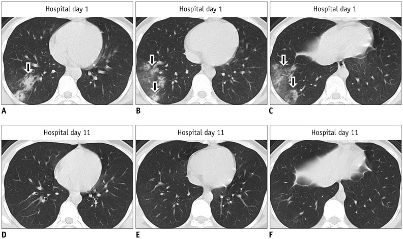
Different slices on same CT scan are horizontally arrayed; same slices on different CT scans are vertically arrayed. A–C. First CT performed on hospital day 1 shows patchy consolidation and ground-glass opacity in right lower lobe (arrows). D–F. Second CT performed on hospital day 11 shows resolution of consolidation and ground-glass opacity. CT = computed tomography
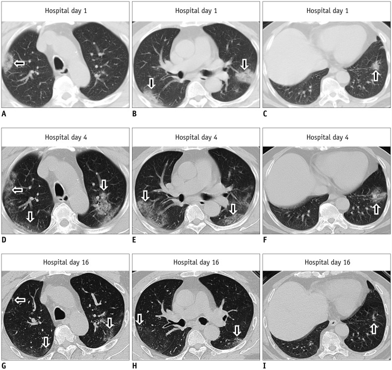
Different slices on same CT scan are horizontally arrayed; same slices on different CT scans are vertically arrayed. A–C. First CT performed on hospital day 1 shows multiple patchy consolidation in subpleural regions of bilateral lungs (arrows). D–F. Second CT performed on hospital day 4 shows decreased consolidation but increased ground-glass opacities in subpleural regions of bilateral lungs (arrows). G–I. Third CT performed on hospital day 16 shows decrease in extent of ground-glass opacities (arrows).
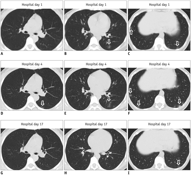
Different slices on same CT scan are horizontally arrayed; same slices on different CT scans are vertically arrayed. A–C. First CT performed on hospital day 1 shows multiple ground-glass opacities in subpleural regions of bilateral lower lobes (arrows). D–F. Second CT performed on hospital day 4 shows increase in ground-glass opacities in subpleural regions of bilateral lower lobes (arrows). G–I. Third CT performed on hospital day 17 shows decrease in ground-glass opacities in subpleural regions of bilateral lower lobes (arrow).
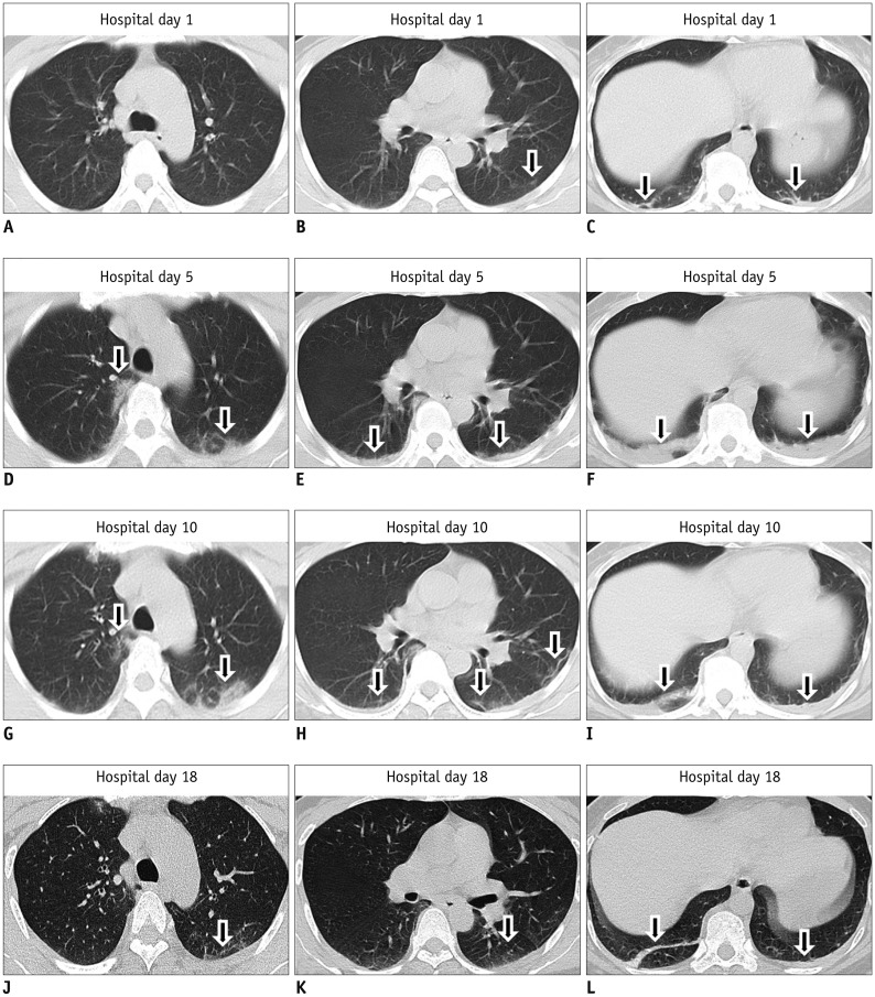
Different slices on same CT scan are horizontally arrayed; same slices on different CT scans are vertically arrayed. A–C. First CT performed on hospital day 1 shows subtle ground-glass opacity and consolidation in subpleural regions of bilateral lungs (arrows). D–F. Second CT performed on hospital day 5 shows increase in ground-glass opacities and consolidation in subpleural regions of bilateral lungs (arrows). G–I. Third CT performed on hospital day 10 shows increase in ground-glass opacities but decrease in consolidation in subpleural regions of bilateral lungs (arrows). J–L. Fourth CT performed on hospital day 18 shows decrease in ground-glass opacities and consolidation (arrows).
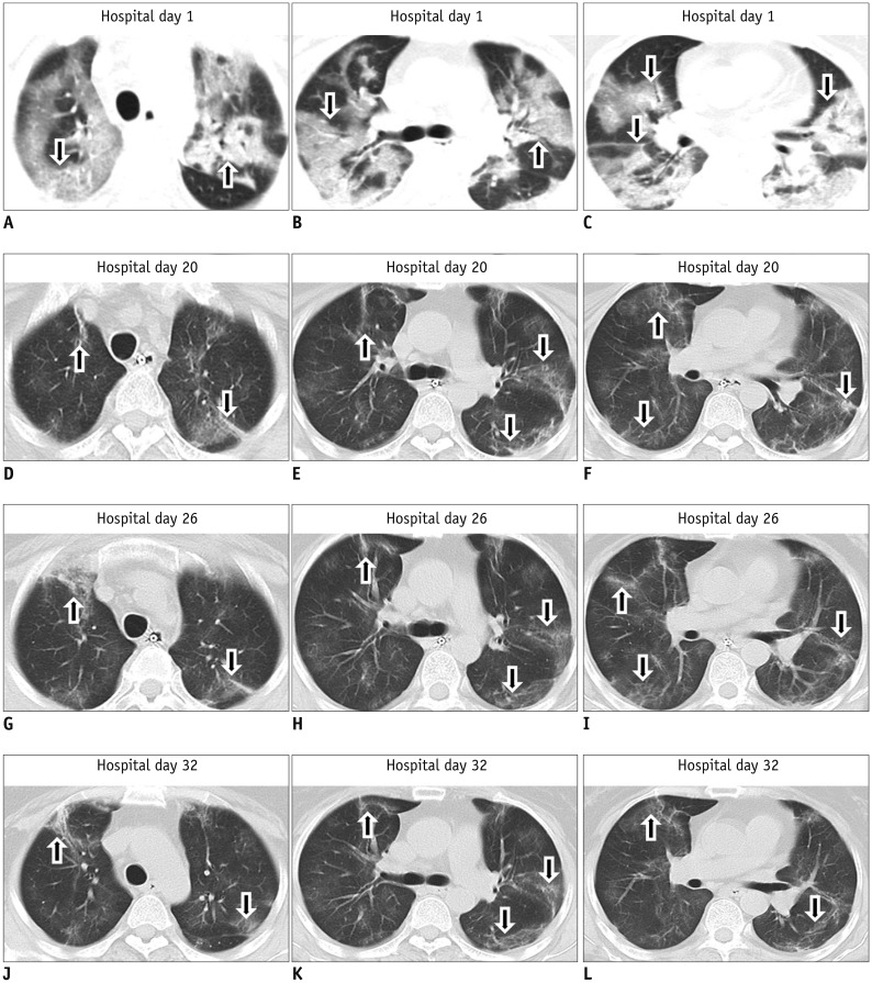
Different slices on same CT scan are horizontally arrayed; same slices on different CT scans are vertically arrayed. A–C. First CT performed on hospital day 1 shows multiple patchy ground-glass opacities and consolidation in bilateral lungs (arrows). D–F. Second CT performed on hospital day 20 shows decrease in ground-glass opacities and consolidation in bilateral lungs (arrows). G–I. Third CT performed on hospital day 26 shows no change in extent of ground-glass opacities and consolidation in bilateral lungs (arrows). J–L. Fourth CT performed on hospital day 32 shows no change in extent of ground-glass opacities and consolidation in bilateral lungs (arrows).
Comment on
-
Novel Coronavirus Pneumonia Outbreak in 2019: Computed Tomographic Findings in Two Cases.Korean J Radiol. 2020 Mar;21(3):365-368. doi: 10.3348/kjr.2020.0078. Epub 2020 Feb 11. Korean J Radiol. 2020. PMID: 32056397 Free PMC article.
References
-
- Clinical management of severe acute respiratory infection when novel coronavirus (nCoV) infection is suspected, interim guidance. World Health Organization Web site. [Accessed February 15, 2020]. https://www.who.int/publications-detail/clinical-management-of-severe-ac.... Published January 12, 2020.
-
- Tian HY. [2019-nCoV: new challenges from coronavirus] Zhonghua Yu Fang Yi Xue Za Zhi. 2020;54:E001. - PubMed
-
- By 24:00 on February 15, 2020, the latest developments of coronavirus disease 2019. National Health Commission of the People's Republic of China Web site. [Accessed February 16, 2020]. http://www.nhc.gov.cn. Published February 16, 2020.
Publication types
MeSH terms
LinkOut - more resources
Full Text Sources
