. 2020 May 1;318(5):G931-G945.
doi: 10.1152/ajpgi.00266.2019. Epub 2020 Mar 16.

Helicobacter pylori infection impairs chaperone-assisted maturation of Na-K-ATPase in gastric epithelium

Affiliations

Helicobacter pylori infection impairs chaperone-assisted maturation of Na-K-ATPase in gastric epithelium

Elizabeth A Marcus et al. Am J Physiol Gastrointest Liver Physiol. .

Abstract

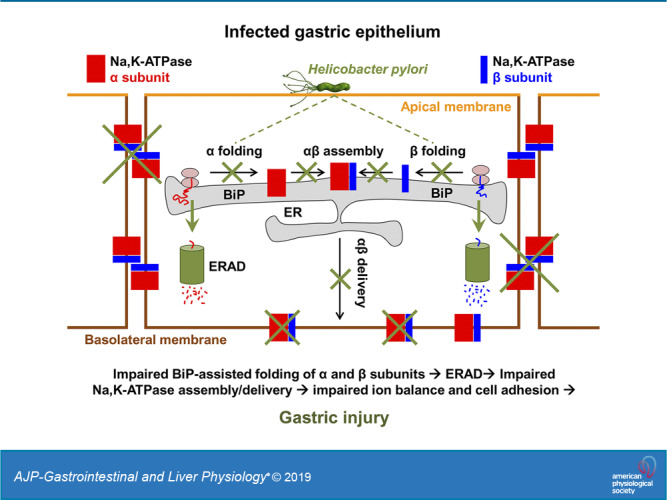
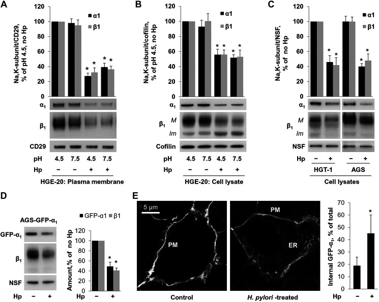
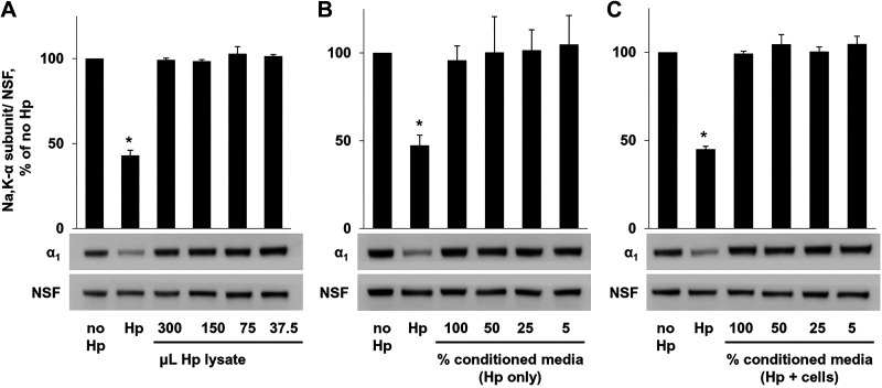
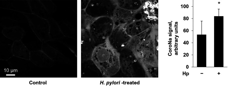
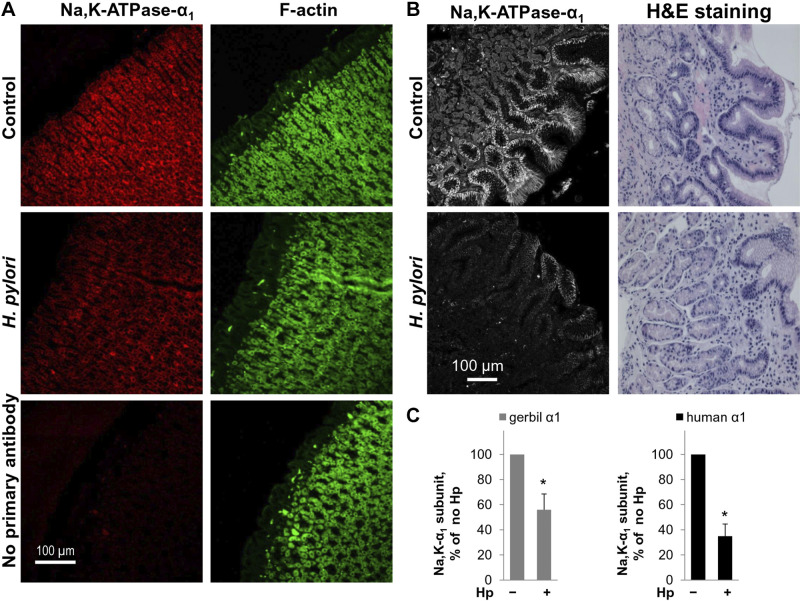
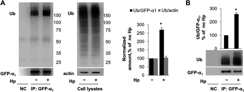
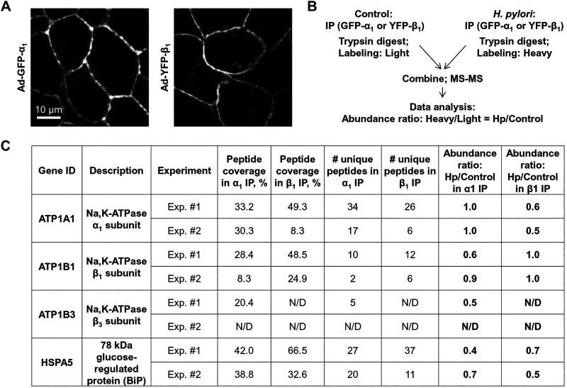
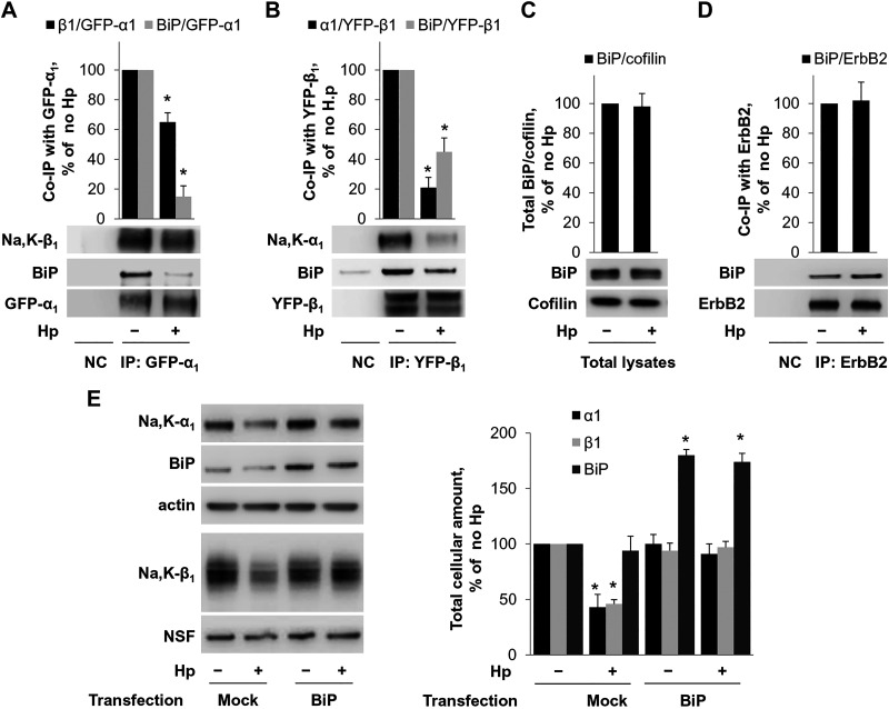
Helicobacter pylori infection always induces gastritis, which may progress to ulcer disease or cancer. The mechanisms underlying mucosal injury by the bacteria are incompletely understood. Here, we identify a novel pathway for H. pylori-induced gastric injury, the impairment of maturation of the essential transport enzyme and cell adhesion molecule, Na-K-ATPase. Na-K-ATPase comprises α- and β-subunits that assemble in the endoplasmic reticulum (ER) before trafficking to the plasma membrane. Attachment of H. pylori to gastric epithelial cells increased Na-K-ATPase ubiquitylation, decreased its surface and total levels, and impaired ion balance. H. pylori did not alter degradation of plasmalemma-resident Na-K-ATPase subunits or their mRNA levels. Infection decreased association of α- and β-subunits with ER chaperone BiP and impaired assembly of α/β-heterodimers, as was revealed by quantitative mass spectrometry and immunoblotting of immunoprecipitated complexes. The total level of BiP was not altered, and the decrease in interaction with BiP was not observed for other BiP client proteins. The H. pylori-induced decrease in Na-K-ATPase was prevented by BiP overexpression, stopping protein synthesis, or inhibiting proteasomal, but not lysosomal, protein degradation. The results indicate that H. pylori impairs chaperone-assisted maturation of newly made Na-K-ATPase subunits in the ER independently of a generalized ER stress and induces their ubiquitylation and proteasomal degradation. The decrease in Na-K-ATPase levels is also seen in vivo in the stomachs of gerbils and chronically infected children. Further understanding of H. pylori-induced Na-K-ATPase degradation will provide insights for protection against advanced disease.NEW & NOTEWORTHY This work provides evidence that Helicobacter pylori decreases levels of Na-K-ATPase, a vital transport enzyme, in gastric epithelia, both in acutely infected cultured cells and in chronically infected patients and animals. The bacteria interfere with BiP-assisted folding of newly-made Na-K-ATPase subunits in the endoplasmic reticulum, accelerating their ubiquitylation and proteasomal degradation and decreasing efficiency of the assembly of native enzyme. Decreased Na-K-ATPase expression contributes to H. pylori-induced gastric injury.

Keywords: Helicobacter pylori; Na-K-ATPase; endoplasmic reticulum; gastric epithelium; protein maturation.


Conflict of interest statement

No conflicts of interest, financial or otherwise, are declared by the author(s).

Figures

None
Graphical abstract
Fig. 1.
Fig. 1.
Helicobacter pylori (Hp) attachment to gastric epithelial cells decreases plasma membrane and total levels of Na-K-ATPase α1- and β1-subunits. HGE-20 cells grown on transwell inserts were infected with H. pylori. Apical pH was adjusted as indicated. A and B: cells were biotinylated on the basolateral side before cell lysis, basolateral surface proteins were extracted using streptavidin beads, and both surface proteins and total cell lysates were analyzed by Western blot. The amount of α1- and β1-subunits of Na-K-ATPase was decreased in the presence of H. pylori independently of pH, in the basolateral membranes (A) and in total cell lysates (B). CD29 was used as loading control for plasma membrane proteins, and cofilin was used as loading control for total cellular proteins. C: results from 2 other gastric cell lines (HGT-1 and AGS) demonstrate a similar decrease in total Na-K-ATPase levels. N-ethylmaleimide-sensitive fusion protein (NSF) was used as a loading control. M, mature fraction; Im, immature fraction. A stable cell line expressing green fluorescent protein (GFP)-α1 was infected with H. pylori. D: Western blot of total cell lysates demonstrated a decrease in the exogenous α1 in the presence of H. pylori. For A–D, representative blots and densitometry quantification results of 3 parallel experiments (means ± SD) are shown. Confocal microscopy demonstrates increased endoplasmic reticulum retention of GFP-α1. E: representative images and quantification of 12 microscopic fields per condition (means ± SD) are shown. ER, endoplasmic reticulum; PM, plasma membrane. *Significant difference from control (no Hp), P < 0.05, t test.
Fig. 2.
Fig. 2.
Attachment of Helicobacter pylori (Hp) to gastric epithelial cells is required for decrease in Na-K-ATPase. A: serial dilutions of H. pylori cell lysate were added to AGS cells, followed by cell lysis and Western blot. No change in Na-K-ATPase was seen with addition of bacterial lysate. Conditioned medium taken from bacterial cultures (B) or from cells infected with H. pylori (C) was applied to AGS cells, followed by lysis and Western blot. Again, no difference was seen in the presence of conditioned medium. Representative blots and densitometry quantification results of 3 parallel experiments (means ± SD) are shown. Controls were vehicle (no Hp) and intact bacteria (Hp). NSF, N-ethylmaleimide-sensitive fusion protein loading control. *Significant difference from control (no Hp), P < 0.05, t test.
Fig. 3.
Fig. 3.
Attachment of Helicobacter pylori (Hp) to gastric cells increases intracellular sodium concentration. HGE-20 cells were incubated for 16 h with or without H. pylori and then loaded with sodium dye coroNa Green-AM. Cells were viewed using confocal microscopy. In the presence of H. pylori, with fewer transporters on the membrane, intracellular sodium was increased compared with control. Experiments were done in triplicate, and fluorescence values represent the average of 10 full fields for each condition. Fluorescence is expressed as means ± SD. *Significant difference from control (no Hp), P < 0.95, t test.
Fig. 4.
Fig. 4.
Helicobacter pylori (Hp) induces decreased expression of Na-K-ATPase in chronic infection. A: gerbils were infected with H. pylori by oral gavage and euthanized after 5 mo. Stomachs were fixed and infection confirmed by Giemsa stain. Immunofluorescence was completed using antibodies against Na-K-ATPase-α1 with F-actin counterstain. B: antrum biopsy samples from pediatric patients with or without H. pylori infection (n = 3 per group) were acquired retrospectively following an IRB-approved pathology database search. Hematoxylin-eosin (H&E) staining was completed (right). Immunofluorescence was performed using antibodies against the α1-subunit of the Na-K-ATPase. Images (left) were obtained by confocal microscopy. C: quantification of 10 microscopic fields for each model is shown as means ± SD), confirming decreased expression of Na-K-ATPase in chronic infection. *Significant difference from control (no Hp), P < 0.05, t test.
Fig. 5.
Fig. 5.
Helicobacter pylori (Hp) induces proteasomal degradation of newly-made Na-K-ATPase subunits. AGS cells (A) or AGS cells stably expressing exogenous green fluorescent protein (GFP)-α1 (B) were incubated with and without H. pylori and inhibitors of lysosomal degradation, bafilomycin (Baf) or CA074-me (CA), or inhibitor of proteosomal degradation lactacystin (Lact), followed by Western blot. V, vehicle. Lactacystin was protective against the effect of H. pylori on both endogenous (A) and exogenous (B) Na-K-ATPase, whereas the other inhibitors had no effect, suggesting a proteasomal degradation pathway. C: HGE-20 cells were biotinylated and then infected with H. pylori for 16 h, and Western blot of the extracted membrane fraction showed no change in rate of degradation of biotinylated Na-K-ATPase subunits in the presence of H. pylori, demonstrating mature transporters are unaffected. D: HGE-20 cells were incubated with and without H. pylori and protein synthesis inhibitor cycloheximide (CHX), followed by lysis and Western blot. CHX prevented the effect of H. pylori on Na-K-ATPase, indicating that H. pylori affects newly made protein. Representative blots and densitometry are shown. NSF (N-ethylmaleimide sensitive fusion protein), ErbB2, and cofilin, loading controls; n = 3 experiments, *Significant difference from control, P < 0.05, t test.
Fig. 6.
Fig. 6.
Helicobacter pylori (Hp) induces ubiquitylation of Na-K-ATPase. A: green fluorescent protein (GFP)-α1 was expressed in HGE-20 cells using an adenovirus vector, followed by immunoprecipitation (IP) using GFP antibodies and Western blot with ubiquitin (Ub) antibodies. H. pylori increased the amount of ubiquitylated forms of the α1-subunit 2.7-fold, whereas total cellular protein ubiquitylation was unaffected. Actin was used as a loading control in total cell lysates. B: ubiquitylation of GFP-α1 stably expressed in AGS cells was also increased as shown. Representative blots and densitometry quantification results of 3 parallel experiments (means ± SD) are shown. *Significant difference from control (no Hp), P < 0.05, t test. Ub, ubiquitin; NC, nontransfected control.
Fig. 7.
Fig. 7.
Exposure of gastric cells to Helicobacter pylori decreases association of Na-K-ATPase subunits with endoplasmic reticulum (ER) chaperone BiP and with each other. A: to enable effective immunoprecipitation (IP) of Na-K-ATPase subunits, green fluorescent protein (GFP)-tagged α1-subunit and yellow fluorescent protein (YFP)-tagged β1-subunit were expressed in HGE-20 cells as confirmed by confocal microscopy. After infection with H. pylori, IP of the subunits was performed using GFP antibodies, and α1- or β1-coimmunoprecipitated proteins were subjected to quantitative mass spectrometry (MS) with dimethyl labeling. B: schema of the experiment is shown. In both α1- and β1-imunoprecipitates, MS results show decreased abundance of ER chaperone BiP and the respective partner Na-K-ATPase subunit(s) in the presence of H. pylori. C: results of 2 independent experiments for each α1- and β1-immunoprecipitate are shown.
Fig. 8.
Fig. 8.
Helicobacter pylori (Hp) decreases Na-K-ATPase by disrupting the Na-K-ATPase-BiP interaction. A and B: yellow fluorescent protein (YFP)-tagged β1-subunit or green fluorescent protein (GFP)-tagged α1-subunit was incorporated into HGE-20 cells and then immunoprecipitated with GFP antibody followed by Western blot, confirming the decrease in association of Na-K-ATPase subunits with BiP and with each other in the presence of H. pylori. C: there is no change in total cellular levels of BiP in the presence of H. pylori, confirming the response is specific to the interaction with the Na-K-ATPase subunits rather than decreased cellular levels of BiP. Cofilin was used as a loading control. D: ErbB2, another plasma membrane protein undergoing folding in the endoplasmic reticulum, does not change association with BiP in the presence of H. pylori. BiP was overexpressed in HGT-1 cells using pcDNA3.1(+)-GRP78/BiP. E: overexpression of BiP prevented the effect of H. pylori on Na-K-ATPase subunits. Representative blots and densitometry quantification results of 3 parallel experiments (means ± SD) are shown for all experiments. NSF (N-ethylmaleimide sensitive fusion protein), loading control. *Significant difference from control (no Hp), P < 0.05, t test. NC, nontransfected control.

Similar articles

Cited by

References

    1. Abdel-Latif AA, Smith JP, Hedrick N. Adenosinetriphosphatase and nucleotide metabolism in synaptosomes of rat brain. J Neurochem 17: 391–401, 1970. doi:10.1111/j.1471-4159.1970.tb02226.x. - DOI - PubMed
    1. Ackermann U, Geering K. Mutual dependence of Na-K-ATPase alpha- and beta-subunits for correct posttranslational processing and intracellular transport. FEBS Lett 269: 105–108, 1990. doi:10.1016/0014-5793(90)81130-G. - DOI - PubMed
    1. Baltrus DA, Amieva MR, Covacci A, Lowe TM, Merrell DS, Ottemann KM, Stein M, Salama NR, Guillemin K. The complete genome sequence of Helicobacter pylori strain G27. J Bacteriol 191: 447–448, 2009. doi:10.1128/JB.01416-08. - DOI - PMC - PubMed
    1. Beggah A, Mathews P, Beguin P, Geering K. Degradation and endoplasmic reticulum retention of unassembled alpha- and beta-subunits of Na,K-ATPase correlate with interaction of BiP. J Biol Chem 271: 20895–20902, 1996. doi:10.1074/jbc.271.34.20895. - DOI - PubMed
    1. Beggah AT, Geering K. Alpha and beta subunits of Na-K-ATPase interact with BiP and calnexin. Ann N Y Acad Sci 834: 537–539, 1997. doi:10.1111/j.1749-6632.1997.tb52311.x. - DOI - PubMed

Publication types

MeSH terms

Substances