. 2020 Jan-Dec:29:963689720910095.
doi: 10.1177/0963689720910095.

In Vivo and In Vitro Evaluation of a Novel Hyaluronic Acid-Laminin Hydrogel as Luminal Filler and Carrier System for Genetically Engineered Schwann Cells in Critical Gap Length Tubular Peripheral Nerve Graft in Rats

Affiliations

In Vivo and In Vitro Evaluation of a Novel Hyaluronic Acid-Laminin Hydrogel as Luminal Filler and Carrier System for Genetically Engineered Schwann Cells in Critical Gap Length Tubular Peripheral Nerve Graft in Rats

Nina Dietzmeyer et al. Cell Transplant. 2020 Jan-Dec.

Abstract

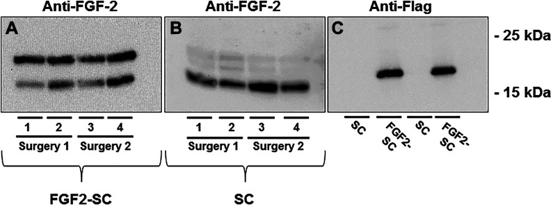
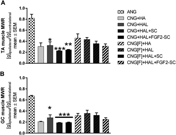
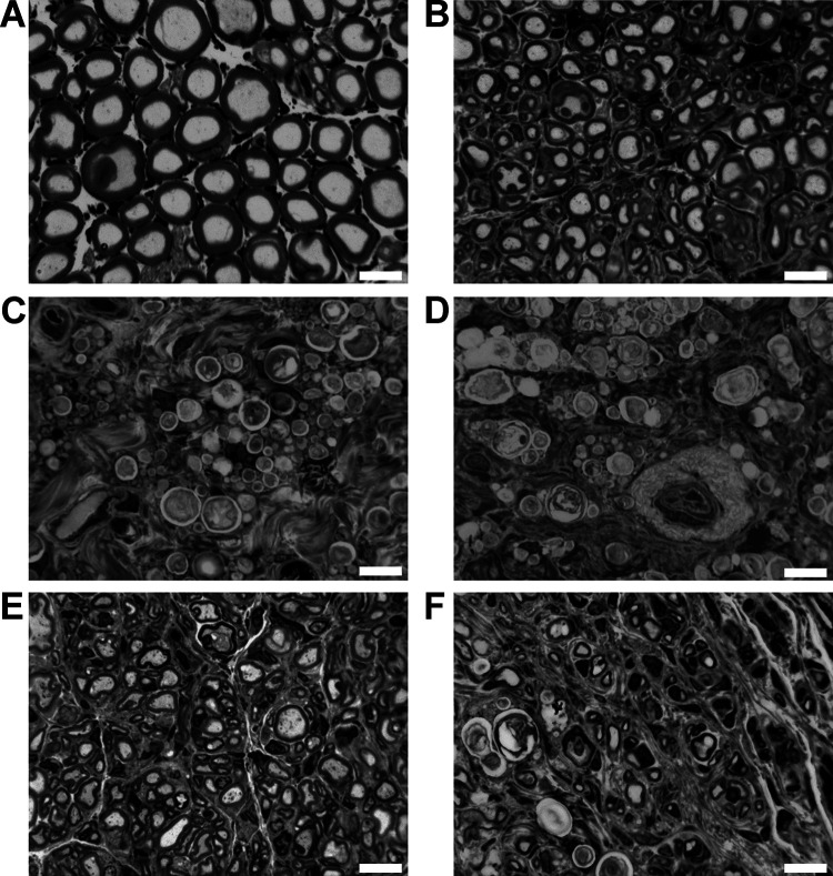
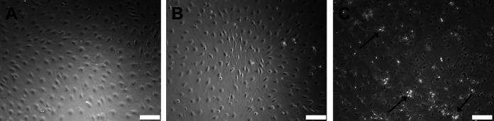
In the current study we investigated the suitability of a novel hyaluronic acid-laminin hydrogel (HAL) as luminal filler and carrier system for co-transplanted cells within a composite chitosan-based nerve graft (CNG) in a rat critical nerve defect model. The HAL was meant to improve the performance of our artificial nerve guides by giving additional structural and molecular support to regrowing axons. We filled hollow CNGs or two-chambered nerve guides with an inserted longitudinal chitosan film (CNG[F]s), with cell-free HAL or cell-free HA or additionally suspended either naïve Schwann cells (SCs) or fibroblast growth factor 2-overexpressing Schwann cells (FGF2-SCs) within the gels. We subjected female Lewis rats to immediate 15 mm sciatic nerve gap reconstruction and comprehensively compared axonal and functional regeneration parameters with the gold standard autologous nerve graft (ANG) repair. Motor recovery was surveyed by means of electrodiagnostic measurements at 60, 90, and 120 days post-reconstruction. Upon explantation after 120 days, lower limb target muscles were harvested for calculation of muscle-weight ratios. Semi-thin cross-sections of nerve segments distal to the grafts were evaluated histomorphometrically. After 120 days of recovery, only ANG treatment led to full motor recovery. Surprisingly, regeneration outcomes revealed no regeneration-supportive effect of HAL alone and even an impairment of peripheral nerve regeneration when combined with SCs and FGF2-SCs. Furthermore, complementary in vitro studies, conducted to elucidate the reason for this unexpected negative result, revealed that SCs and FGF2-SCs suspended within the hydrogel relatively downregulated gene expression of regeneration-supporting neurotrophic factors. In conclusion, cell-free HAL in its current formulation did not qualify for optimizing regeneration outcome through CNG[F]s. In addition, we demonstrate that our HAL, when used as a carrier system for co-transplanted SCs, changed their gene expression profile and deteriorated the pro-regenerative milieu within the nerve guides.

Keywords: Schwann cells; cellular drug delivery system; chitosan; fibroblast growth factor 2; sciatic nerve regeneration.


Conflict of interest statement

Declaration of Conflicting Interests: The author(s) declared no potential conflicts of interest with respect to the research, authorship, and/or publication of this article.

Figures

Fig 1.
Fig 1.
Results of western blot analyses of cell lysates from naïve or genetically engineered Schwann cells. Detection of endogenous fibroblast growth factor 2 (FGF-2) expression (A and B) and Flag-tagged FGF-218kDA overexpression (C) and by western blotting of cell lysates derived from naïve Schwann cells (SCs) and genetically engineered FGF-2-overexpressing Schwann cells (FGF2-SCs) cultured for 24 h in specific SC medium and another 24 h in serum-free N2 medium. Endogenous FGF-218 kDa and FGF-223 kDa were detected in lysates of FGF2-SCs, showing a strong signal for both isoforms. In naïve SCs all isoforms of FGF-2 (18 kDA, 21 kDa, and 23 kDa) were detected with a strong signal only detected for FGF-218 kDa. FGF-218kDA-Flag was detected in lysates derived from FGF2-SCs, while lysates of naïve SCs did not result in any detection. Each n = 4 (indicated by columns 1 to 4).
Fig 2.
Fig 2.
Bar graphs depicting the muscle weight ratios (MWRs) of tibialis anterior (A, TA muscle) and gastrocnemius (B, GC muscle) muscles at 120 days after reconstruction surgery. Kruskal–Wallis test with Dunn’s multiple comparisons were applied to detect significant differences (*P < 0.05, **P < 0.01, ***P < 0.001 vs. autologous nerve graft [ANG]). Results are presented as mean ± SEM.
Fig 3.
Fig 3.
Macroscopical appearance of the regenerated tissue between the proximal and the distal nerve end upon tissue harvest 120 days after reconstruction. (A) Macroscopic appearance of an autologous nerve graft (ANG), this registered in category 1 = substantial tissue regeneration. (B) Another example for category 1 regeneration, here from the CNG+HAL group. (C) An example of category 3 tissue regeneration, a hair thin connection between the two nerve ends, here from the CNG+HAL+FGF2-SC group. (D) Tissue regeneration through two-chambered CNG[F]s in some cases resulted in the formation of two tissue cables also registered in category 1. The example is derived from the CNG[F]+HAL group and additionally demonstrates vascularized tissue bridges that have grown through the perforations in the chitosan film (arrows).
Fig 4.
Fig 4.
Representative pictures of toluidine blue-stained semi-thin cross-sections of distal nerve segments 120 days after reconstruction surgery. Images show healthy nerve segments (A), serving as control compared to distal nerve segments of reconstructed sciatic nerves (B–F). Examples for no axonal regeneration from (C) CNG+HAL group and (D) the CNG+HAL+fibroblast growth factor 2 (FGF2)-Schwann cell (SC) group. Examples of samples, demonstrating axonal regeneration from (B) the autologous nerve graft group, (E) the CNG[F]+HAL group, and (F) the CNG[F]+HAL+FGF2-SC group. White scale bars display 10 µm.
Fig 5.
Fig 5.
Representative pictures of phase-contrast microscopy of Schwann cells (SC) seeded in either SC-specific culture medium (K+, A), hyaluronic acid (HA, B), and hyaluronic acid–laminin hydrogel (HAL, C). Three days after seeding, cells cultured in K+ (A) and HA (B) showed a typical bipolar morphology. Proliferation of the initially seeded 350,000 cells led to a dense cell layer on the well ground. In HAL condition (C), SC, however, revealed a different morphology and higher apoptosis rate (cell detritus is indicated by arrows). Scale bar displays 100 µm.
Fig 6.
Fig 6.
Gene expression changes in naïve Schwann cells (SCs, A-D) and fibroblast growth factor 2 (FGF-2)-overexpressing Schwann cells (FGF2-SC, E-H). Gene expression of bdnf, gdnf, ngf, and fgf-2 was quantified by qRT-PCR of cDNA of SCs and FGF2-SCs cultured in either specific SC medium (K+), hyaluronic acid (HA), or hyaluronic acid–laminin hydrogel (HAL). Bdnf is significantly higher expressed in HA-cultured FGF2-SCs when compared to K+-cultured FGF2-SCs (E). Relative amounts of transcripts to the housekeeping gene ppia are shown as mean ± SD. One-way analysis of variance followed by Dunnett’s multiple comparison were applied to detect significant differences (*P < 0.05 vs. K+). Naïve SCs in HAL: n = 2, rest: n = 3.

Similar articles

Cited by

References

    1. Asplund M, Nilsson M, Jacobsson A, von Holst H. Incidence of traumatic peripheral nerve injuries and amputations in Sweden between 1998 and 2006. Neuroepidemiology. 2009;32(3):217–228. - PubMed
    1. Noble J, Munro CA, Prasad VS, Midha R. Analysis of upper and lower extremity peripheral nerve injuries in a population of patients with multiple injuries. J Trauma. 1998;45(1):116–122. - PubMed
    1. Eriksson M, Karlsson J, Carlsson KS, Dahlin LB, Rosberg HE. Economic consequences of accidents to hands and forearms by log splitters and circular saws: cost of illness study. J Plast Surg Hand Surg. 2011;45(1):28–34. - PubMed
    1. Deumens R, Bozkurt A, Meek MF, Marcus MA, Joosten EA, Weis J, Brook GA. Repairing injured peripheral nerves: bridging the gap. Prog Neurobiol. 2010;2010(3):245–276. - PubMed
    1. Grinsell D, Keating CP. Peripheral nerve reconstruction after injury: a review of clinical and experimental therapies. Biomed Res Int.2014;2014:698256. - PMC - PubMed

Publication types

LinkOut - more resources