Ultrasonographic assessment of airway
- PMID: 32174650
- PMCID: PMC7047677
- DOI: 10.4103/joacp.JOACP_319_18
Ultrasonographic assessment of airway
Abstract
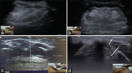
Ultrasound is gaining increasing popularity among anesthesiologists as it is readily available and provides real-time imaging for various procedures. It is considered as a "visual stethoscope" of the anesthesiologist. After establishing its use in regional blocks and central venous catheter insertion, it is now finding increasing use in anticipation of difficult airway and securing and maintaining it. It has challenged the classical approach of clinical assessment of airway and allows more dynamic bedside assessment. This article attempts to briefly outline the role of ultrasound and its applications for airway management in patients.
Keywords: Airway; anesthesia; clinical; sonographic; ultrasound.
Copyright: © 2020 Journal of Anaesthesiology Clinical Pharmacology.
Conflict of interest statement
There are no conflicts of interest.
Figures


References
-
- Singh M, Chin KJ, Chan VWS, Wong DT, Prasad GA, Yu E. Use of sonography for airway assessment: An observational study. J Ultrasound Med Off J Am Inst Ultrasound Med. 2010;29:79–85. - PubMed
-
- Lun H-M, Zhu S-Y, Liu R-C, Gong J-G, Liu Y-L. Investigation of the upper airway anatomy with ultrasound. Ultrasound Q. 2016;32:86–92. - PubMed
-
- Valente T, Farina R, Minelli S, Pinto A, Rossi G, Tecame S, et al. [The echographic anatomy of the larynx and the perilaryngeal structures] Radiol Med (Torino) 1996;91:231–7. - PubMed