. 2020 Feb 27:14:1.
doi: 10.3389/fncir.2020.00001. eCollection 2020.

Fictive Scratching Patterns in Brain Cortex-Ablated, Midcollicular Decerebrate, and Spinal Cats

Affiliations

Fictive Scratching Patterns in Brain Cortex-Ablated, Midcollicular Decerebrate, and Spinal Cats

Irene Guadalupe Aguilar Garcia et al. Front Neural Circuits. .

Abstract

Background: The spinal cord's central pattern generators (CPGs) have been explained by the symmetrical half-center hypothesis, the bursts generator, computational models, and more recently by connectome circuits. Asymmetrical models, at odds with the half-center paradigm, are composed of extensor and flexor CPG modules. Other models include not only flexor and extensor motoneurons but also motoneuron pools controlling biarticular muscles. It is unknown whether a preferred model can explain some particularities that fictive scratching (FS) in the cat presents. The first aim of this study was to investigate FS patterns considering the aiming and the rhythmic periods, and second, to examine the effects of serotonin (5HT) on and segmental inputs to FS. Methods: The experiments were carried out first in brain cortex-ablated cats (BCAC), then spinalized (SC), and for the midcollicular (MCC) preparation. Subjects were immobilized and the peripheral nerves were used to elicit the Monosynaptic reflex (MR), to modify the scratching patterns and for electroneurogram recordings. Results: In BCAC, FS was produced by pinna stimulation and, in some cases, by serotonin. The scratching aiming phase (AP) initiates with the activation of either flexor or extensor motoneurons. Serotonin application during the AP produced simultaneous extensor and flexor bursts. Furthermore, WAY 100635 (5HT1A antagonist) produced a brief burst in the tibialis anterior (TA) nerve, followed by a reduction in its electroneurogram (ENG), while the soleus ENG remained silent. In SC, rhythmic phase (RP) activity was recorded in the soleus motoneurons. Serotonin or WAY produced FS bouts. The electrical stimulation of Ia afferent fibers produced heteronymous MRes waxing and waning during the scratch cycle. In MCC, FS began with flexor activity. Electrical stimulation of either deep peroneus (DP) or superficial peroneus (SP) nerves increased the duration of the TA electroneurogram. Medial gastrocnemius (MG) stretching or MG nerve electrical stimulation produced a reduction in the TA electroneurogram and an initial MG extensor burst. MRes waxed and waned during the scratch cycle. Conclusion: Descending pathways and segmental afferent fibers, as well as 5-HT and WAY, can change the FS pattern. To our understanding, the half-center hypothesis is the most suitable for explaining the AP in MCC.

Keywords: decerebrate cat; fictive scratching; monosynaptic reflex; motor patterns; spinal cat.


Figures

Figure 1
Figure 1
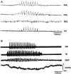
Short episodes of fictive scratching (FS) appeared after d-tubocurarine application with two characteristics: absence of SOL electroneurographic activity and DC shifts in spinal cord intermediate nucleus. (A) Traces: flexor hallucis longus (FHL), lateral gastrocnemius (LG), flexor digitorum longus (FDL) and soleus (SOL). The ENGs were integrated and rectified. (B) DC potential during FS scratching. Traces: first, gastrocnemius soleus (GS); second, tibial anterior (TA). Traces 1 and 2 are raw electroneurograms. Third trace: dorsal root potential (DRP), fourth trace: DC potential shift.
Figure 2
Figure 2
(A) FS produced by pinna stimulation and bursting activity with a slower frequency in SOL ENG; FS was rectified and integrated ENG in FHL, FDL and SOL ENG exhibits a slow frequency rhythm produced by FP mechanical stimulation. Traces as indicated. (B) Slow frequency spontaneous rhythmic activity in the MG ENG. After pinna stimulation, slow frequency rhythmic activity concurred with FS frequency in the MG ENG. The red arrow indicates FP stimulation.
Figure 3
Figure 3
Heteronymous Monosynaptic reflex (MR) modulation in brain cortex-ablated cats (BCAC) during the rhythmic phase (RP) of the FS. (A) HeMR amplitude changes in FHL motoneurons (ordinate), produced by electrical MG nerve stimulation (S-MG) plotted in relation to the normalized FHL scratch cycle (abscissa). Upper bar below the abscissa (SC) denotes cycle duration as measured in the FHL ENG. Second and third bars below the abscissa are the extensor and flexor phase durations, respectively. The dot with the vertical line on the left of the ordinate indicates the HeMR amplitude and standard deviation (SD) before the scratching episode. (B–D) HeMR amplitude changes in ST, LG, and FHL motoneurons in relation to normalized ST, LG, and FHL scratch cycles, respectively. Electrical stimulation was given to the PB nerve (1.3 T). In B, ST-HeMR amplitude changes (ordinate) in relation to the ST scratch cycle (abscissa). The continuous line in panel (B) represents the averaged and rectified ST ENG. The extensor phase is indicated. The mean scratching cycle duration and its SD were measured in the ST ENG. Asterisk in (A–D) denotes P < 0.05. In (C,D), the HeMR was recorded in LG and FHL nerves, respectively. Other indications as in (A).
Figure 4
Figure 4
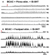
FS episode elicited by pinna stimulation plus 5HT in a BCAC. (A) Pinna stimulation without 5HT (traces as indicated MG, TA, and PB). (B) After brainstem 5HT microinjection indicated with an arrow (traces as indicated PB, MG, and TA). The traces are consecutive recordings. In (B), the ENG activity before RP onset occurred in PB and MG. The red arrow indicates pinna stimulation.
Figure 5
Figure 5
Different FS patterns in BCAC produced by pinna stimulation plus 5HT. (A) The AP commences with flexor activity in the TA ENG. (B) FS in a different cat after spinal cord 5HT microinjection. The AP commences with ENG activity in the MG and PB ENG. (C) FS episode showing tonic AP with simultaneous activity on flexor and extensor ENG. FS episodes (A–C) were recorded in three different cats. ENGs, integrated and rectified. The red arrow indicates burst activity in MG and PB, ENG before TA-ENG activity during the AP.
Figure 6
Figure 6
FS episode elicited by pinna stimulation in a BCAC and SC after intraspinal cord 5HT microinjection. (A) BCAC, traces as indicated (TA, MG, and SOL). (B) SC, traces as indicated (TA, MG, and SOL), note the scratching SOL ENG activity. ENGs rectified and integrated. (C) The graph insert shows the peak amplitude means values of TA, MG, and SOL, P < 0.05.
Figure 7
Figure 7
FS and fictive locomotion (FL) evoked in a BCAC by pinna stimulation and mechanical FP stimulation with 5HT applied in the spinal cord. (A) An episode of FS evoked by pinna stimulation in a 5HT-treated cat. The red arrow indicates pinna stimulation. (B) An episode of FL evoked by FP stimulation in a 5HT-treated cat. The red arrow indicates FP stimulation.
Figure 8
Figure 8
(A) ENGs of eight hindlimb nerves during an episode of FS evoked by pinna stimulation after WAY-100635 microinjection in the L4 spinal cord in a BCAC. Traces as indicated (TA, MG, FDL, PB, FHL, LG, ST, and PER). (B) FS in a different cat produced by pinna stimulation and WAY-100635 in the L5 segment in the spinal cord of a BCAC. Traces as indicated (TA, FHL, GM, and PB). Note the TA ENG activity at the onset of the FS episode, also a sustained reduction in TA ENG during the RP. The red arrows indicates pinna stimulation with a burst in the TA-ENG.
Figure 9
Figure 9
(A) FS in spinal cat after 5HT microinjection. Traces as indicated (TA, FDL, SOL, and PB). 5HT produces rhythmic ENG activity in the SOL nerve. (B) FS in an SC after microinjection of WAY-100635. Traces as indicated (TA, GM, and SOL). EMG traces were integrated and rectified.
Figure 10
Figure 10
FS bouts in spinal cats produced by WAY-100635, FS commences with long duration bursts in FDL and PB ENGs (A), with a burst in FHL (B), or short bursts in TA, FDL, and MG nerves (C). ENG amplitude varied for different bouts of FS (TA, FDL, MG, and PB). EMG traces were integrated and rectified. The red arrow indicates pinna stimulation and the FS episode begun with a short burst of activity in TA, FDL, MG nerves.
Figure 11
Figure 11
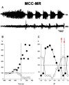
(A) An episode of FS in an MCC recorded in the TA and MG nerves. (B) Normalized amplitudes of MR responses before and during the approach period in a single experiment. AP onset is indicated by the vertical interrupted line at time zero. The reflex is produced by single electrical shocks applied to the sectioned L7 roots at variable intervals before or after the beginning of pinna stimulation. Black dots, TA; white dots, MG; MR amplitude values. (C) Normalized MR amplitude during the rhythmic period. 100% value indicated by a hatched line in the ordinate (note the logarithmic scale in ordinates of C). Abscissa normalized cycle. The values between hatched lines correspond to MG MR activity. Between red arrows indicate the extensor phase of the scratch cycle.
Figure 12
Figure 12
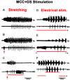
Effects produced by activation of afferent fibers from extensor muscles in the FS pattern. Nerve activity was recorded from the TA nerve and a small filament in the MG nerve. Panels (A–C), (D–F), two different experiments. Panels (A,D) are control ENGs, traces as indicated (TA and MG). In (B,C), the triceps surae muscle continuously stretched 3 mm (B), 6 mm (C); the stretch was applied approximately 3 s before pinna stimulation. Note in (B,C) the shortening of the TA approach period and the pre-extensor discharges; also note in (C) the prolonged extensor firing and the interruption of the RP despite continuous pinna stimulation. In (D–F), the lower trace (S) indicates the time of respective electrical stimulation (300 Hz) of the MG filament at 3.1 times the threshold of the most excitable afferent fibers. In (E), the activities commence with extensor activity that terminated although nerve stimulation was maintained. Note in (F), electrical stimulation applied during the RP increased the MG burst duration 7- to 8-fold. The red arrow indicates EMG, ENG burst produces by triceps surae stretching, 3 or 6 mm respectively.
Figure 13
Figure 13
Effects produced by superficial or deep peroneus (DP) nerve electrical stimulation in the FS pattern. (A,B) Electrical stimulation to superficial peroneus (SP) prolongs the TA AP. (C,D) The DP nerve stimulation prolongs the TA burst with a corresponding decrease in MG activity. When nerve stimulation coincides with TA activity, the duration of TA bursts increases; when the stimulus begins during MG activity, the TA post-discharge also increases but in a minor proportion (arrow).

Similar articles

References

    1. Aggelopoulos N. C., Burton M. J., Clarke R. W., Edgley S. A. (1996). Characterization of a descending system that enables crossed group II inhibitory reflex pathways in the cat spinal cord. J. Neurosci. 16, 723–729. 10.1523/jneurosci.16-02-00723.1996 - DOI - PMC - PubMed
    1. Alaburda A. (2005). Periodic high-conductance states in spinal neurons during scratch-like network activity in adult turtles. J. Neurosci. 25, 6316–6321. 10.1523/jneurosci.0843-05.2005 - DOI - PMC - PubMed
    1. Anderssson O., Grillner S. (1981). Peripheral control of the cat’s step cycle I. Phase dependent effects of ramp-movements of the hip during “fictive locomotion”. Acta Physiol. Scand. 113, 89–101. 10.1111/j.1748-1716.1981.tb06867.x - DOI - PubMed
    1. Ausborn J., Shevtsova N. A., Caggiano V., Danner S. M., Rybak I. A. (2019). Computational modeling of brainstem circuits controlling locomotor frequency and gait. Elife 8:e43587. 10.7554/elife.43587 - DOI - PMC - PubMed
    1. Ausborn J., Snyder A. C., Shevtsova N. A., Rybak I. A., Rubin J. E. (2018). State-dependent rhythmogenesis and frequency control in a half-center locomotor CPG. J. Neurophysiol. 119, 96–117. 10.1152/jn.00550.2017 - DOI - PMC - PubMed

MeSH terms