

Enhanced Phenotype Definition for Precision Isolation of Precursor Exhausted Tumor-Infiltrating CD8 T Cells
- PMID: 32174925
- PMCID: PMC7056729
- DOI: 10.3389/fimmu.2020.00340
Enhanced Phenotype Definition for Precision Isolation of Precursor Exhausted Tumor-Infiltrating CD8 T Cells
Abstract
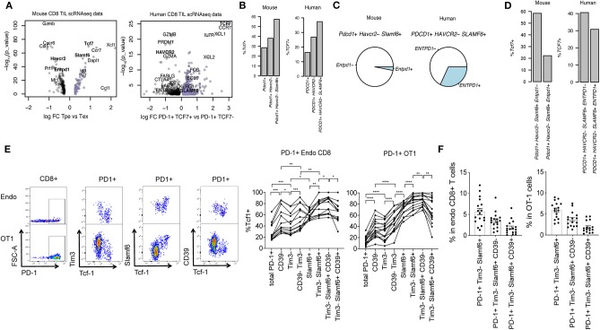
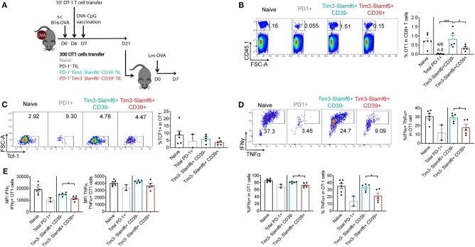
In the context of adoptive T cell transfer (ACT) for cancer treatment, it is crucial to generate in vitro large amounts of tumor-specific CD8 T cells with high potential to persist in vivo. PD-1, Tim3, and CD39 have been proposed as markers of tumor-specific tumor-infiltrating CD8 T lymphocytes (CD8 TILs). However, these molecules are highly expressed by terminally differentiated exhausted CD8 T cells (Tex) that lack proliferation potential. Therefore, optimized strategies to isolate tumor-specific TILs with high proliferative potential, such as Tcf1+ precursor exhausted T cells (Tpe) are needed to improve in vivo persistence of ACT. Here we aimed at defining cell surface markers that would unequivocally identify Types for precision cell sorting increasing the purity of tumor-specific PD-1+ Tcf1+ Tpe from total TILs. Transcriptomic analysis of Tpe vs. Tex CD8 TIL subsets from B16 tumors and primary human melanoma tumors revealed that Tpes are enriched in Slamf6 and lack Entpd1 and Havcr2 expression, which encode Slamf6, CD39, and Tim3 cell surface proteins, respectively. Indeed, we observed by flow cytometry that CD39- Tim3- Slamf6+ PD-1+ cells yielded maximum enrichment for tumor specific PD-1+ Tcf1+ OT1 TILs in B16.OVA tumors. Moreover, this population showed higher re-expansion capacity upon an acute infection recall response compared to the CD39+ counterparts or bulk PD-1+ TILs. Hence, we report an enhanced sorting strategy (CD39- Tim3- Slamf6+ PD-1+) of Tpes. In conclusion, we show that optimization of CD8 TIL cell sorting strategy is a viable approach to improve recall capacity and in vivo persistence of transferred cells in the context of ACT.
Keywords: adoptive cell transfer; melanoma; memory-like CD8 T cells; precursor exhausted T cells; tumor-infiltrating CD8 T cells.
Copyright © 2020 Martinez-Usatorre, Carmona, Godfroid, Yacoub Maroun, Labiano and Romero.
Figures
Similar articles
-
Intratumoral Tcf1+PD-1+CD8+ T Cells with Stem-like Properties Promote Tumor Control in Response to Vaccination and Checkpoint Blockade Immunotherapy.Immunity. 2019 Jan 15;50(1):195-211.e10. doi: 10.1016/j.immuni.2018.12.021. Epub 2019 Jan 8. Immunity. 2019. PMID: 30635237
-
Stem-like CD8 T cells mediate response of adoptive cell immunotherapy against human cancer.Science. 2020 Dec 11;370(6522):1328-1334. doi: 10.1126/science.abb9847. Science. 2020. PMID: 33303615 Free PMC article.
-
Deciphering the transcriptomic landscape of tumor-infiltrating CD8 lymphocytes in B16 melanoma tumors with single-cell RNA-Seq.Oncoimmunology. 2020 Mar 12;9(1):1737369. doi: 10.1080/2162402X.2020.1737369. eCollection 2020. Oncoimmunology. 2020. PMID: 32313720 Free PMC article.
-
Adoptive T-cell therapy using autologous tumor-infiltrating lymphocytes for metastatic melanoma: current status and future outlook.Cancer J. 2012 Mar-Apr;18(2):160-75. doi: 10.1097/PPO.0b013e31824d4465. Cancer J. 2012. PMID: 22453018 Free PMC article. Review.
-
Technology meets TILs: Deciphering T cell function in the -omics era.Cancer Cell. 2023 Jan 9;41(1):41-57. doi: 10.1016/j.ccell.2022.09.011. Epub 2022 Oct 6. Cancer Cell. 2023. PMID: 36206755 Free PMC article. Review.
Cited by
-
CXCR5+CD8 T cells: Potential immunotherapy targets or drivers of immune-mediated adverse events?Front Med (Lausanne). 2022 Oct 13;9:1034764. doi: 10.3389/fmed.2022.1034764. eCollection 2022. Front Med (Lausanne). 2022. PMID: 36314014 Free PMC article. Review.
-
Tumor-infiltrating lymphocytes in the immunotherapy era.Cell Mol Immunol. 2021 Apr;18(4):842-859. doi: 10.1038/s41423-020-00565-9. Epub 2020 Nov 2. Cell Mol Immunol. 2021. PMID: 33139907 Free PMC article. Review.
-
Charged Particle and Conventional Radiotherapy: Current Implications as Partner for Immunotherapy.Cancers (Basel). 2021 Mar 23;13(6):1468. doi: 10.3390/cancers13061468. Cancers (Basel). 2021. PMID: 33806808 Free PMC article. Review.
-
Combination of multiple omics techniques for a personalized therapy or treatment selection.Front Immunol. 2023 Sep 27;14:1258013. doi: 10.3389/fimmu.2023.1258013. eCollection 2023. Front Immunol. 2023. PMID: 37828984 Free PMC article. Review.
-
Solid Tumor Microenvironment Can Harbor and Support Functional Properties of Memory T Cells.Front Immunol. 2021 Nov 11;12:706150. doi: 10.3389/fimmu.2021.706150. eCollection 2021. Front Immunol. 2021. PMID: 34867942 Free PMC article.
References
Publication types
MeSH terms
Substances
LinkOut - more resources
Full Text Sources
Research Materials