

Acacetin Attenuates Renal Damage-Induced by Ischemia-Reperfusion with Declining Apoptosis and Oxidative Stress in Mice
- PMID: 32175062
- PMCID: PMC7050221
- DOI: 10.4103/ijpvm.IJPVM_512_18
Acacetin Attenuates Renal Damage-Induced by Ischemia-Reperfusion with Declining Apoptosis and Oxidative Stress in Mice
Abstract
Background: Renal ischemia-reperfusion disturbs both the function and the histology of this organ. Acacetin (Aca) is a natural flavonoid that is effective for relief of many diseases. The aim of this study was to determine the impacts of Aca on renal ischemia-reperfusion process in mice.
Methods: In total, 84 male Balb/cmice divided into 12 groups and were administrated intraperitoneally for 4 days with or without surgery to dimethyl sulfoxide 0.01% or Aca (10, 25, and 50 mg/kg) as Control, control Acas, sham, sham Acas groups. Ischemia-reperfusion without or with Aca (10, 25, and 50 mg/kg) treatments were the other groups. Parameters related to the function and the histology of the kidneys were evaluated and statistically analyzed from kidney and blood serum samples in the respect of the groups.
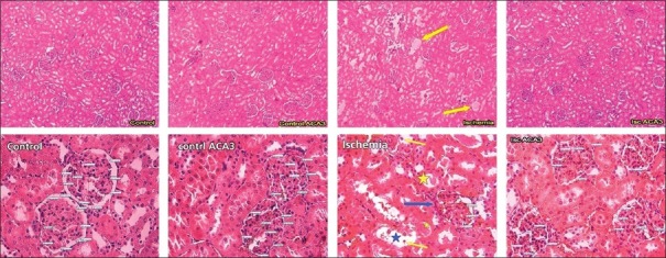
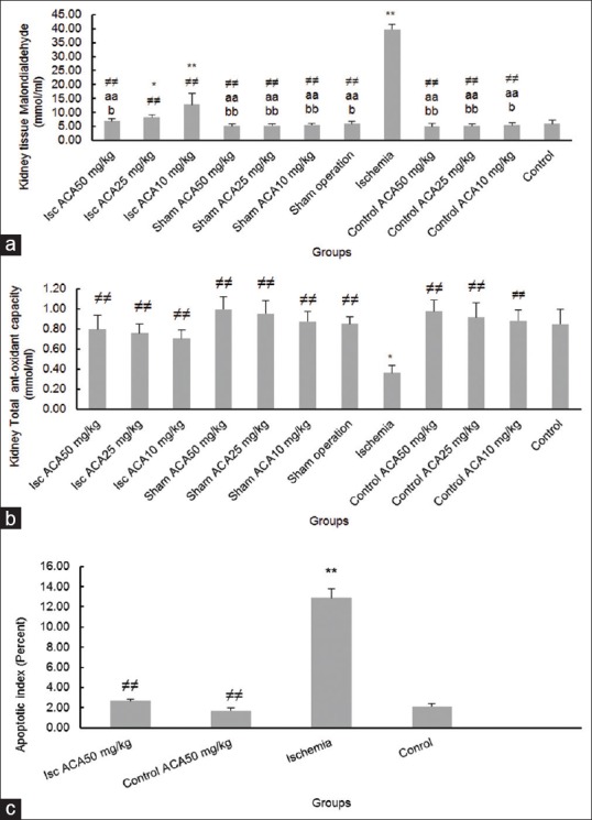
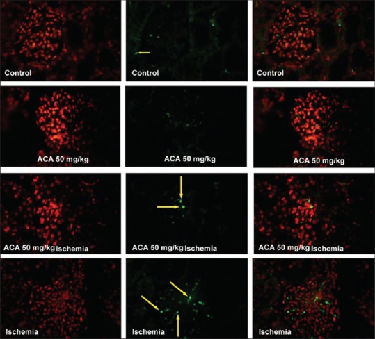
Results: In ischemia-reperfusion and ischemia-reperfusion + Aca (10 mg/kg) groups, there were significantly increased in urea, creatinine, malondialdehyde (MDA), and apoptosis rate, whereas total antioxidant capacity decreased compared to the control and sham and ischemia-reperfusion + Aca (25 and 50 mg/kg) (P < 0.05). The histopathology alteration was seen in the ischemia-reperfusion group than the others (P < 0.01). Moreover, there was a significant difference between ischemia-reperfusion + Aca (25 and50 mg/kg) groups than ischemia-reperfusion + Aca (10 mg/kg) one (P < 0.05).
Conclusions: The recovery effect of Aca was offered on renal ischemia-reperfusion damage in a dose-dependent manner in mice, showing by kidney histopathology and functional criteria improvements. The attributed mechanism for this impression would be the antioxidant property of Aca, decreasing both MDA levels and apoptosis rate in kidney tissue.
Keywords: Acacetin; antioxidants; apoptosis; malondialdehyde; reperfusion injury.
Copyright: © 2020 International Journal of Preventive Medicine.
Conflict of interest statement
There are no conflicts of interest.
Figures
Similar articles
-
Acacetin Alleviates Hepatitis Following Renal Ischemia-Reperfusion in Male Balb/C Mice by Antioxidants Regulation and Inflammatory Markers Suppression.J Invest Surg. 2021 May;34(5):495-503. doi: 10.1080/08941939.2019.1656309. Epub 2019 Nov 5. J Invest Surg. 2021. PMID: 31686554
-
Protective effect of N-(p-amylcinnamoyl) anthranilic acid, phospholipase A2 enzyme inhibitor, and transient receptor potential melastatin-2 channel blocker against renal ischemia-reperfusion injury.J Cell Biochem. 2019 Mar;120(3):3822-3832. doi: 10.1002/jcb.27664. Epub 2018 Sep 27. J Cell Biochem. 2019. PMID: 30259992
-
Anti-inflammatory and antioxidant activity of essential amino acid α-ketoacid analogues against renal ischemia-reperfusion damage in Wistar rats.Biomedica. 2020 Jun 15;40(2):336-348. doi: 10.7705/biomedica.4875. Biomedica. 2020. PMID: 32673461 Free PMC article.
-
ERK phosphorylation plays an important role in the protection afforded by hypothermia against renal ischemia-reperfusion injury.Surgery. 2017 Feb;161(2):444-452. doi: 10.1016/j.surg.2016.07.028. Epub 2016 Aug 31. Surgery. 2017. PMID: 27590616
-
Protective effect of tea polyphenols on renal ischemia/reperfusion injury via suppressing the activation of TLR4/NF-κB p65 signal pathway.Gene. 2014 May 25;542(1):46-51. doi: 10.1016/j.gene.2014.03.021. Epub 2014 Mar 12. Gene. 2014. PMID: 24630969
Cited by
-
Effect of acacetin on inhibition of apoptosis in Helicobacter pylori-infected gastric epithelial cell line.World J Gastrointest Oncol. 2024 Aug 15;16(8):3624-3634. doi: 10.4251/wjgo.v16.i8.3624. World J Gastrointest Oncol. 2024. PMID: 39171164 Free PMC article.
-
Acacetin as a natural cardiovascular therapeutic: mechanisms and preclinical evidence.Front Pharmacol. 2025 Apr 4;16:1493981. doi: 10.3389/fphar.2025.1493981. eCollection 2025. Front Pharmacol. 2025. PMID: 40255574 Free PMC article. Review.
-
RNA Sequencing Analyses Reveal the Potential Anti-Inflammatory Mechanisms of Acacetin Against ODG/R Injuries in Microglia.J Inflamm Res. 2024 Jun 5;17:3641-3652. doi: 10.2147/JIR.S465093. eCollection 2024. J Inflamm Res. 2024. PMID: 38855167 Free PMC article.
-
Acacetin alleviates neuroinflammation and oxidative stress injury via the Nrf2/HO-1 pathway in a mouse model of spinal cord injury.Transl Neurosci. 2022 Dec 21;13(1):483-494. doi: 10.1515/tnsci-2022-0266. eCollection 2022 Jan 1. Transl Neurosci. 2022. PMID: 36590896 Free PMC article.
-
Harmine protects mercuric chloride kidney-induced injury by antioxidant activity in male mice: a biochemical and histological study.Res Pharm Sci. 2020 Nov 27;15(6):541-550. doi: 10.4103/1735-5362.301339. eCollection 2020 Dec. Res Pharm Sci. 2020. PMID: 33828597 Free PMC article.
References
-
- Volpe A, Blute ML, Ficarra V, Gill IS, Kutikov A, Porpiglia F, et al. Renal ischemia and function after partial nephrectomy: A collaborative review of the literature. Eur Urol. 2015;68:61–74. - PubMed
-
- Janero DR. Malondialdehyde and thiobarbituric acid-reactivity as diagnostic indices of lipid peroxidation and peroxidative tissue injury. Free Radic Biol Med. 1 99;;9:515–40. - PubMed
-
- Rodrigo R, Bosco C. Oxidative stress and protective effects of polyphenols: Comparative studies in human and rodent kidney. A review. Comp Biochem PhysiolC: Toxicol Pharmacol. 2006;142:317–27. - PubMed
LinkOut - more resources
Full Text Sources