. 2020 Feb;8(4):114.
doi: 10.21037/atm.2019.12.88.

Tumor slice culture as a biologic surrogate of human cancer

Affiliations

Tumor slice culture as a biologic surrogate of human cancer

Heidi L Kenerson et al. Ann Transl Med. 2020 Feb.

Abstract

Background: The tumor microenvironment (TME) is critical to every aspect of cancer biology. Organotypic tumor slice cultures (TSCs) preserve the original TME and have demonstrated utility in predicting drug sensitivity, but the association between clinicopathologic parameters and in vitro TSC behavior has not been well-defined.

Methods: One hundred and eight fresh tumor specimens from liver resections at a tertiary academic center were procured and precisely cut with a Vibratome to create 250 μm × 6 mm slices. These fixed-dimension TSCs were grown on polytetrafluoroethylene inserts, and their metabolic activities were determined by a colorimetric assay. Correlation between baseline activities and clinicopathologic parameters was assessed. Tissue CEA mRNA expression was determined by RNAseq.

Results: By standardizing the dimensions of a slice, we found that adjacent tumor slices have equivalent metabolic activities, while those derived from different tumors exhibit >30-fold range in baseline MTS absorbances, which correlated significantly with the percentage of tumor necrosis based on histologic assessment. Extending this to individual cancers, we were able to detect intra-tumoral heterogeneity over a span of a few millimeters, which reflects differences in tumor cell density and Ki-67 positivity. For colorectal cancers, tissue CEA expression based on RNAseq of tumor slices was found to correlate with clinical response to chemotherapies.

Conclusions: We report a standardized method to assess and compare human cancer growth ex vivo across a wide spectrum of tumor samples. TSC reflects the state of tumor behavior and heterogeneity, thus providing a simple approach to study of human cancers with an intact TME.

Keywords: Organotypic; cancer model; heterogeneity; microenvironment.


Conflict of interest statement

Conflicts of Interest: The authors have no conflicts of interest to declare.

Figures

Figure 1
Figure 1
Standardized assessment of tumor slice cultures (TSCs) by precision cutting. (A) Workflow of the TSC platform from surgical specimen to slices. Left, representative tumor ‘wedge’ removed from surgical specimen; middle, 6-mm core from a punch biopsy; right, agarose-embedded cores set in Vibratome for slicing. Refer to Supplemental Methods for details; (B) comparison of baseline MTS absorbance with and without normalization to wet weight of slices from 2 cases of metastatic colorectal carcinoma (CRC). The table shows the variance of weight and absorbance measurements; (C) baseline MTS absorbance of 3 consecutive TSCs each from 4 different tumors without normalization to wet weight. HCC, hepatocellular carcinoma.
Figure 2
Figure 2
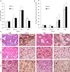
Viability of tumor slices in vitro. Assessment of viability and proliferation by MTS assay (A) and Ki-67 expression (B) in tumor slices maintained in vitro for 7 days compared to baseline (day 0); (C) histologic appearances of the different tumor types based on H&E staining and Ki-67 immunostaining corresponding to samples highlighted in A. CRC, colorectal carcinoma; HCC, hepatocellular carcinoma; ICC, intrahepatic cholangiocarcinoma. Original magnification 200×.
Figure 3
Figure 3
Baseline MTS absorbance of fixed dimension tumor slices from 53 consecutive tumors. Values represent at least 3 biologic replicates taken randomly throughout each tumor. CRC, colorectal carcinoma; HCC, hepatocellular carcinoma; ICC, intrahepatic cholangiocarcinoma; FNH, focal nodular hyperplasia; FLC, fibrolamellar carcinoma.
Figure 4
Figure 4
Baseline MTS value as an indicator of intra-tumoral heterogeneity. (A) Regional heterogeneity: MTS values from tumor cores obtained from 3 different sites within tumors >3 cm from 5 different patients. Each site is at least 1 cm from the others and is representd by at least 3 slices. *, P<0.05 compared to site 1; (B) histologic correlation with baseline MTS from CRC-1 shown in A. Top panels: H&E representing each of the 3 sites; bottom panel: Ki-67 IHC of cores from site 1 and site 3. Even though site 1 is more cellular than site 3, the proportion of Ki-67+ cells is significantly lower than site 3 (site 1: 21% vs. site 3: 82%). All photos are at 40× magnification; (C) local heterogeneity: Each tumor core was sectioned serially along its length and baseline MTS values were determined for each slice. Left column: Examples of tumor cores showing minor heterogeneity (<1 absorbance unit variation). Right column: Examples of tumor cores showing major heterogeneity (>1 absorbance unit variation). Examples of tumor histology indicated by the red circles are shown in two cases of CRC. Tumor cells are highlighted by purple stain. CRC, colorectal carcinoma; HCC, hepatocellular carcinoma; ICC, intrahepatic cholangiocarcinoma.
Figure 5
Figure 5
Tissue mRNA expression of CEA correlates negatively with clinical response to chemotherapies. Tumo slices from metastatic CRC in the liver were analyzed by RNAseq to determine level of expression of CEACAM5 gene that encodes CEA. Results are compared to those of cytokeratin 20 (KRT20) and a housekeeping gene, GAPDH. Response groups are based on clinical data from patients receiving chemotherapies prior the surgical resection.

References

    1. Marquart J, Chen EY, Prasad V. Estimation of the Percentage of US Patients With Cancer Who Benefit From Genome-Driven Oncology. JAMA Oncol 2018;4:1093-8. 10.1001/jamaoncol.2018.1660 - DOI - PMC - PubMed
    1. Arnedos M, Vicier C, Loi S, et al. Precision medicine for metastatic breast cancer--limitations and solutions. Nat Rev Clin Oncol 2015;12:693-704. 10.1038/nrclinonc.2015.123 - DOI - PubMed
    1. Vlachostergios PJ, Faltas BM. Treatment resistance in urothelial carcinoma: an evolutionary perspective. Nat Rev Clin Oncol 2018;15:495-509. 10.1038/s41571-018-0026-y - DOI - PubMed
    1. Morash M, Mitchell H, Beltran H, et al. The Role of Next-Generation Sequencing in Precision Medicine: A Review of Outcomes in Oncology. J Pers Med 2018;8. doi: .10.3390/jpm8030030 - DOI - PMC - PubMed
    1. Hyman DM, Puzanov I, Subbiah V, et al. Vemurafenib in Multiple Nonmelanoma Cancers with BRAF V600 Mutations. N Engl J Med 2015;373:726-36. 10.1056/NEJMoa1502309 - DOI - PMC - PubMed