Review
. 2020 May 1;98(5):skaa081.
doi: 10.1093/jas/skaa081.

Satellite cells and their regulation in livestock

Affiliations
Review

Satellite cells and their regulation in livestock

Madison L Gonzalez et al. J Anim Sci. .

Abstract

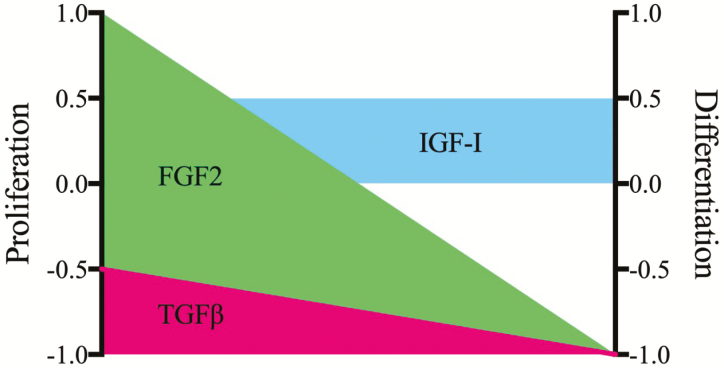
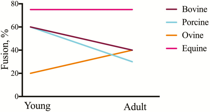
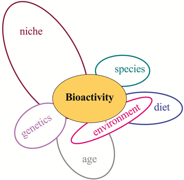
Satellite cells are the myogenic stem and progenitor population found in skeletal muscle. These cells typically reside in a quiescent state until called upon to support repair, regeneration, or muscle growth. The activities of satellite cells are orchestrated by systemic hormones, autocrine and paracrine growth factors, and the composition of the basal lamina of the muscle fiber. Several key intracellular signaling events are initiated in response to changes in the local environment causing exit from quiescence, proliferation, and differentiation. Signals emanating from Notch, wingless-type mouse mammary tumor virus integration site family members, and transforming growth factor-β proteins mediate the reversible exit from growth 0 phase while those initiated by members of the fibroblast growth factor and insulin-like growth factor families direct proliferation and differentiation. Many of these pathways impinge upon the myogenic regulatory factors (MRF), myogenic factor 5, myogenic differentiation factor D, myogenin and MRF4, and the lineage determinate, Paired box 7, to alter transcription and subsequent satellite cell decisions. In the recent past, insight into mouse transgenic models has led to a firm understanding of regulatory events that control satellite cell metabolism and myogenesis. Many of these niche-regulated functions offer subtle differences from their counterparts in livestock pointing to the existence of species-specific controls. The purpose of this review is to examine the mechanisms that mediate large animal satellite cell activity and their relationship to those present in rodents.

Keywords: livestock; myogenesis; paired box 7; satellite cell; skeletal muscle.


Figures

Figure 1.
Figure 1.
Morphological features of a bovine embryo at day 23 of gestation. Embryos were harvested at 23 d of gestation and examined for developmental milestones. Gross morphology of the embryo (A) demonstrating the presence of branchial arches (BA, i), a beating four-chamber heart (H, ii), and the presumptive forelimb bud (PL, iii). Bar represents 1 mm.
Figure 2.
Figure 2.
Myogenesis in the early bovine embryo. Day 23 and 45 bovine embryos were fixed and immunostained for key myogenic muscle proteins. MyHC (A) expression localized to the somites (S) and heart (H) of a day 23 embryo. Cryosections through the rostral somites were immunostained for Pax7 (green) and counterstained with phalloidin (red) (B). Coronal sections through a day 45 embryo stained with Hoechst 33324 for nuclei (C). The red box denotes a forelimb with magnification in the remaining panels. Pax7 and Myf5 co-localize to distinct limb myoblast subpopulations. Bar represents 1 mm or 50 µm.
Figure 3.
Figure 3.
Primary regulators of satellite cell proliferation and differentiation in vitro. Satellite cell isolates from meat-producing livestock cultured in permissive media with the FGF2 (green) elicit a strong proliferative response with a corresponding inhibition of differentiation. Modest mitogenic and myogenic actions are found for IGF-I (blue) with TGF-β (red) serving as a general inhibitor of proliferation and a strong suppressor of differentiation. Scale +1 denotes maximal positive response and −1 maximal inhibitory response.
Figure 4.
Figure 4.
Donor age and species differences impact satellite cell myogenic capacity in vitro. Satellite cells were isolated from young and adult cattle, sheep, pigs, and horses, cultured to confluence and examined at various times after fusion. The extent of myoblast fusion reported by authors (see text) graphed as a function of age. Young is equivalent to pre-weaning and adult is considered post-pubertal and beyond the inflection point on a traditional growth curve.
Figure 5.
Figure 5.
Satellite cell bioactivity is regulated by multiple factors. Summary of the literature provides evidence for multiple intrinsic (genetics, niche, age) and extrinsic factors (diet, environment) that impinge upon one another to affect the decisions of the satellite cell.

References

    1. Adams, G. R., Caiozzo V. J., Haddad F., and Baldwin K. M.. . 2002. Cellular and molecular responses to increased skeletal muscle loading after irradiation. Am. J. Physiol. Cell Physiol. 283:C1182–C1195. doi:10.1152/ajpcell.00173.2002 - DOI - PubMed
    1. Alexander, L. S., Mahajan A., Odle J., Flann K. L., Rhoads R. P., and Stahl C. H.. . 2010. Dietary phosphate restriction decreases stem cell proliferation and subsequent growth potential in neonatal pigs. J. Nutr. 140:477–482. doi:10.3945/jn.109.117390 - DOI - PubMed
    1. Alexander, L. S., Seabolt B. S., Rhoads R. P., and Stahl C. H.. . 2012. Neonatal phosphate nutrition alters in vivo and in vitro satellite cell activity in pigs. Nutrients. 4:436–448. doi:10.3390/nu4060436 - DOI - PMC - PubMed
    1. Allen, R. E., and Boxhorn L. K.. . 1989. Regulation of skeletal muscle satellite cell proliferation and differentiation by transforming growth factor-beta, insulin-like growth factor I, and fibroblast growth factor. J. Cell. Physiol. 138:311–315. doi:10.1002/jcp.1041380213 - DOI - PubMed
    1. Allen, R. E., Sheehan S. M., Taylor R. G., Kendall T. L., and Rice G. M.. . 1995. Hepatocyte growth factor activates quiescent skeletal muscle satellite cells in vitro. J. Cell. Physiol. 165:307–312. doi:10.1002/jcp.1041650211 - DOI - PubMed