

Diagnosis of drowning using postmortem computed tomography combined with endoscopic autopsy: A case report
- PMID: 32176043
- PMCID: PMC7220088
- DOI: 10.1097/MD.0000000000019182
Diagnosis of drowning using postmortem computed tomography combined with endoscopic autopsy: A case report
Abstract
Rationale: Postmortem forensic imaging technologies provide a noninvasive/minimally invasive approach for imaging of internal organ structures of the human body to detect injuries, diseases, and other morphologic changes. Currently, postmortem forensic imaging methods have been widely used in determination of the cause of death. However, these methods do not allow histologic examinations. Endoscopic autopsy emerged in the 1990s. Thoracoscopy and laparoscopy are mainly used to examine organs and tissues in the thoracic and abdominal cavity. Target tissues are also sampled for histologic examination. By combining postmortem forensic imaging with endoscopic autopsy, comprehensive examination of the corpse, organs, and sampling for histologic examination can be carried out.
Patient concerns: A 34-year-old woman was witnessed jumping into the river, sinking after struggling in the water. The body was found 24 hours later and confirmed with no vital signs. No preexisting medical conditions were known.
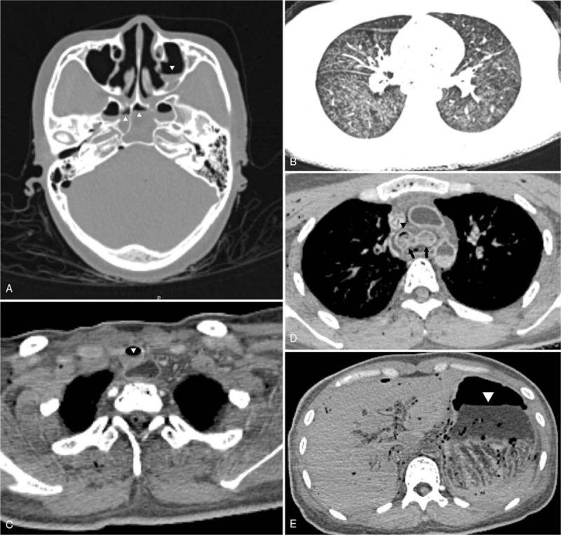
Diagnosis, interventions, and outcomes: Postmortem computed tomography, target coronary postmortem computed tomography angiography, and endoscopic autopsy were performed before conventional autopsy. Laparoscopic examination was used to examine the abdominal organs. The diaphragm and pericardium were cut open from the abdominal cavity to allow access to the examination of lungs and heart. Tissue samples were collected from various organs for histologic examination, and a diatom test was carried out on lung samples. Postmortem computed tomography revealed fluid in the paranasal sinuses, airways, stomach, and duodenum; emphysema aquosum; and mosaic pattern of the lung parenchyma. Endoscopic examination additionally detected Paltauf spots. The results were consistent with those of conventional autopsy. Histologic examination revealed pulmonary congestion, pulmonary edema, pulmonary emphysema, pulmonary hemorrhage, and congestion in multiple organs such as the liver, spleen, and kidneys. Diatoms were detected in lung tissues, which were identical in morphology to diatoms in water samples collected from the scene. The cause of death was determined as drowning.
Conclusion: Combining forensic imaging and endoscopic autopsy for postmortem examination yields a more comprehensive and scientific finding, and the combination is minimally invasive and more acceptable to the family members. This method can be used as an alternative for conventional autopsy under specific circumstances.
Conflict of interest statement
The authors have no conflicts of interest to disclose.
Figures


Similar articles
-
[Postmortem CT examination in a case of alleged drowning--a case report].Arch Med Sadowej Kryminol. 2009 Oct-Dec;59(4):330-6. Arch Med Sadowej Kryminol. 2009. PMID: 20860307 Polish.
-
The number of diatoms recovered from the lungs and other organs in drowning deaths in bathwater.Leg Med (Tokyo). 2011 Jul;13(4):186-90. doi: 10.1016/j.legalmed.2011.04.002. Epub 2011 May 11. Leg Med (Tokyo). 2011. PMID: 21565542
-
Postmortem computed tomography findings in cases of bath-related death: Applicability and limitation in forensic practice.Forensic Sci Int. 2018 Jan;282:195-203. doi: 10.1016/j.forsciint.2017.11.030. Epub 2017 Dec 2. Forensic Sci Int. 2018. PMID: 29223918
-
The persistent problem of drowning - A difficult diagnosis with inconclusive tests.J Forensic Leg Med. 2019 Aug;66:79-85. doi: 10.1016/j.jflm.2019.06.003. Epub 2019 Jun 13. J Forensic Leg Med. 2019. PMID: 31229802 Review.
-
Diagnosis of drowning using post-mortem computed tomography - state of the art.Arch Med Sadowej Kryminol. 2014;64(2):59-75. doi: 10.5114/amsik.2014.47744. Arch Med Sadowej Kryminol. 2014. PMID: 25574940 Review.
Cited by
-
Videoautopsy-A Minimally Invasive Autopsy Method Using Endoscopic Techniques in Forensic Medicine: Clinical Features.Diagnostics (Basel). 2024 Apr 24;14(9):884. doi: 10.3390/diagnostics14090884. Diagnostics (Basel). 2024. PMID: 38732299 Free PMC article.
-
Evaluation of eye and serum findings in different waters in rabbits by drowning and submersion modeling.Turk J Med Sci. 2023 Nov 11;54(1):42-51. doi: 10.55730/1300-0144.5764. eCollection 2024. Turk J Med Sci. 2023. PMID: 38812651 Free PMC article.
-
Minimally invasive autopsy - endoscopic approach to post-mortem diagnostics.Wideochir Inne Tech Maloinwazyjne. 2024 Mar;19(1):122-128. doi: 10.5114/wiitm.2023.134122. Epub 2023 Dec 29. Wideochir Inne Tech Maloinwazyjne. 2024. PMID: 38974768 Free PMC article. Review.
References
-
- Avrahami R, Watemberg S, Daniels-Philips E, et al. Endoscopic autopsy. Am J Forensic Med Pathol 1995;16:147–50. - PubMed
-
- Damore IILJ, Barth RF, Morrison CD, et al. Laparoscopic postmortem examiantion: a minimally invasive approach to the autopsy. Ann Diagn Pathol 2000;4:95–8. - PubMed
-
- Avrahami R, Watemberg S, Hiss Y, et al. Laparoscopic vs conventional autopsy: a promising perspective. Arch Surg 1995;130:407–9. - PubMed
-
- Avrahami R, Watemberg S, Hiss Y. Thoracoscopy vs conventional autopsy of the thorax: a promising perspective. Arch Surg 1995;130:956–8. - PubMed
Publication types
MeSH terms
LinkOut - more resources
Full Text Sources
Medical
