

Chondromyxoid fibroma of the temporal bone: A rare case report
- PMID: 32176085
- PMCID: PMC7440231
- DOI: 10.1097/MD.0000000000019487
Chondromyxoid fibroma of the temporal bone: A rare case report
Abstract
Rationale: Chondromyxoid fibroma (CMF) is a rare form of benign bone tumor and easily misdiagnosed as fibrosarcoma. Hence, to explore the clinical manifestations, diagnostic tests, and therapeutic procedures for temporal bone cartilage myxoid fibroma, it is important to optimize patient treatment and avoid overtreatment. Previous research has discussed cases of CMF, but this paper presents a systematic, complete, and comprehensive introduction of this disease based on this case and related literature.
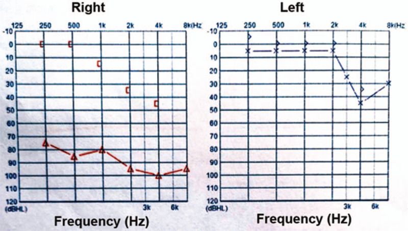
Patient concerns: A 52-year-old male patient presented with pain in his right ear for 2 years and hearing loss in his right ear with tinnitus for 1 year. The patient had a history of hypertension for 9 years and it was well-controlled.
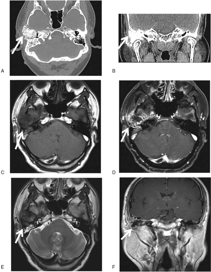
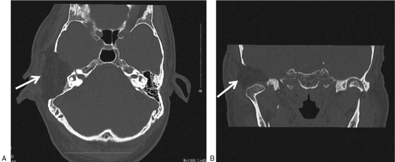
Diagnosis: A computed tomography (CT) scan of the temporal bone showed an expansive growth on the right temporal bone plate and tympanic plate, presenting as a cloud-like ground glass opaque shadow involving the temporom and ibular joint, middle skull base, and small auditory bones. A magnetic resonance imaging (MRI) of the temporal bone showed a large and irregular soft tissue mass shadow on the right temporal bone plate. The right temporal bone plate was occupied by the lesion, consistent with a bone origin. From the results of the imaging examination of the patient, a lesion occupying the temporal bone in the right ear and mastoiditis in the right middle ear was initially diagnosed.
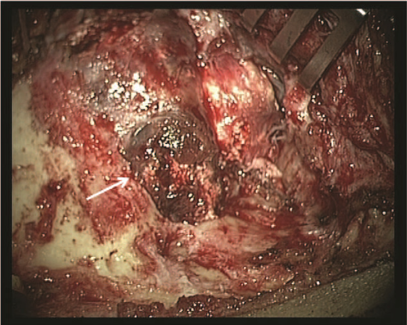
Interventions: Right ear temporal bone tumor resection and abdominal fat extraction were conducted.
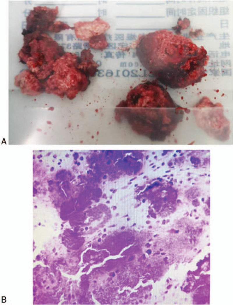
Outcomes: Postoperative pathological results demonstrated myxoid fibroma of the temporal bone cartilage. No recurrence or severe complications were observed in 8 months of follow-up.
Lessons: A finding of myxoid fibroma of the temporal bone cartilage is rare in the clinic. The growth of such tumors is slow. The temporal bone CT and inner ear MRI were helpful in diagnosis. Surgery was the principal treatment.
Conflict of interest statement
The authors have no conflicts of interest to disclose.
Figures
References
-
- Jaffe HL, Lichtenstein L. Chondromyxoid fibroma of bone; a distinctive benign tumor likely to be mistaken especially for chondrosarcoma. Arch Pathol (Chic) 1948;45:541–51. - PubMed
-
- Lingen MW, Solt DB, Polverini PJ. Unusual presentation of a chondromyxoid fibroma of the mandible. Report of a case and review of the literature. Oral Surg Oral Med Oral Pathol 1993;75:615–21. - PubMed
-
- Lang S, Adler CP. Bone diseases: macroscopic, histological and radiological diagnosis of structural changes in skeleton, Springer-Verlag, 2000, pages 589, DM 269, ISBN 3-540-65061-X. Eur J Radiol 2001;37:139.
Publication types
MeSH terms
LinkOut - more resources
Full Text Sources
