. 2021 Jan 1;117(1):201-211.
doi: 10.1093/cvr/cvaa066.

Pim1 maintains telomere length in mouse cardiomyocytes by inhibiting TGFβ signalling

Affiliations

Pim1 maintains telomere length in mouse cardiomyocytes by inhibiting TGFβ signalling

David E Ebeid et al. Cardiovasc Res. .

Abstract

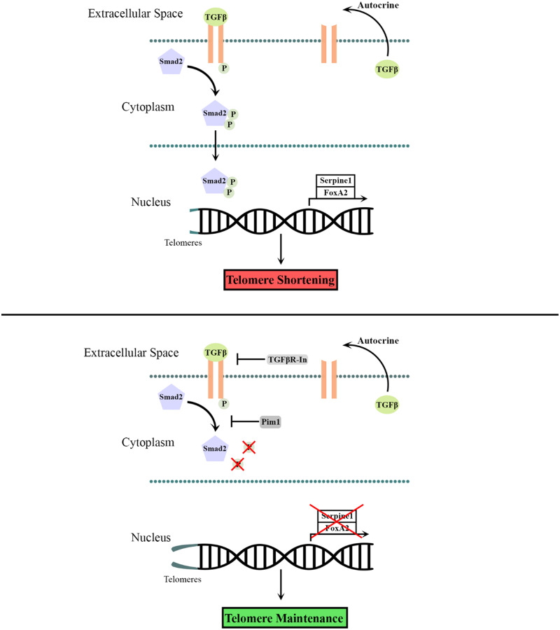
Aims: Telomere attrition in cardiomyocytes is associated with decreased contractility, cellular senescence, and up-regulation of proapoptotic transcription factors. Pim1 is a cardioprotective kinase that antagonizes the aging phenotype of cardiomyocytes and delays cellular senescence by maintaining telomere length, but the mechanism remains unknown. Another pathway responsible for regulating telomere length is the transforming growth factor beta (TGFβ) signalling pathway where inhibiting TGFβ signalling maintains telomere length. The relationship between Pim1 and TGFβ has not been explored. This study delineates the mechanism of telomere length regulation by the interplay between Pim1 and components of TGFβ signalling pathways in proliferating A549 cells and post-mitotic cardiomyocytes.

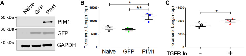
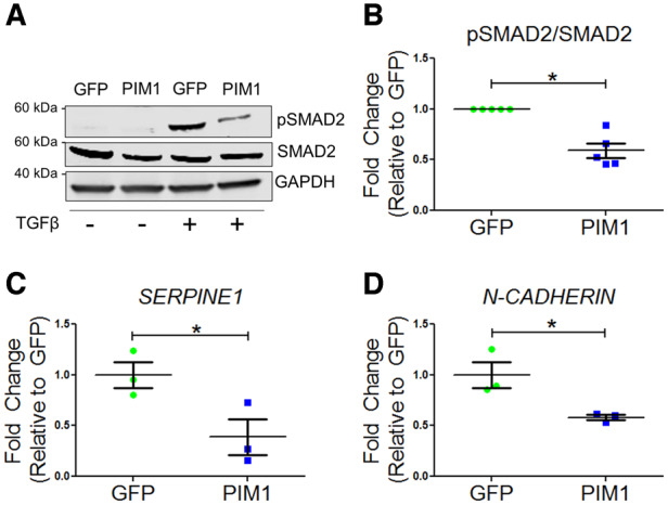
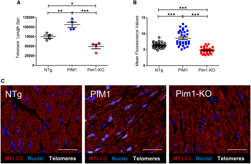
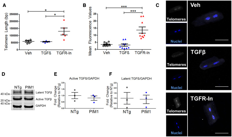
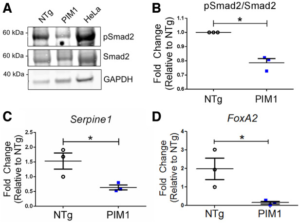
Methods and results: Telomere length was maintained by lentiviral-mediated overexpression of PIM1 and inhibition of TGFβ signalling in A549 cells. Telomere length maintenance was further demonstrated in isolated cardiomyocytes from mice with cardiac-specific overexpression of PIM1 and by pharmacological inhibition of TGFβ signalling. Mechanistically, Pim1 inhibited phosphorylation of Smad2, preventing its translocation into the nucleus and repressing expression of TGFβ pathway genes.

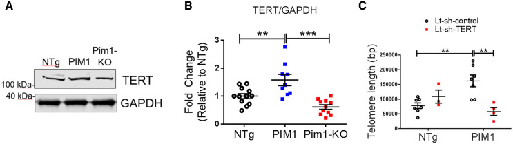
Conclusion: Pim1 maintains telomere lengths in cardiomyocytes by inhibiting phosphorylation of the TGFβ pathway downstream effectors Smad2 and Smad3, which prevents repression of telomerase reverse transcriptase. Findings from this study demonstrate a novel mechanism of telomere length maintenance and provide a potential target for preserving cardiac function.

Keywords: Cardiomyocyte; Pim1; Smad2; TGFβ; Telomere.


Figures

None
Graphical abstract
Figure 1
Figure 1
Telomere length is maintained by PIM1 overexpression or TGFβ receptor inhibition. (A) Representative western-blot bands of GFP and PIM1 in A549 cells transduced with GFP or PIM1-GFP. (B) qRT–PCR quantification of telomere lengths in transduced A549 cells. n = 3, error bars represent SEM, **P < 0.01 vs. GFP, *P < 0.05 vs. naive as measured by one-way ANOVA followed by Tukey’s multiple comparison test. (C) qRT–PCR quantification of telomere lengths in naive A549 cells treated with TGFR-In. n = 4, error bars represent SEM, *P < 0.05 vs. untreated as measured by Student’s t-test.
Figure 2
Figure 2
TGFβ signalling is blunted by PIM1. (A) Representative western blot of pSMAD2 and total SMAD2 in GFP and PIM1 cells with and without treatment of 4 ng/mL TGFβ. (B) Quantification of pSMAD2 over total SMAD2. n = 5, error bars represent SEM, *P < 0.01 vs. GFP as measured by Student’s t-test. (C) Quantification of SERPINE1 and (D) N-CADHERIN mRNA expression via qRT–PCR. n = 3, error bars represent SEM, *P < 0.05 vs. GFP as measured by Student’s t-test.
Figure 3
Figure 3
Telomere length is maintained by PIM1 in adult murine cardiomyocytes. (A) Analysis of telomere lengths in isolated adult cardiomyocytes from PIM1 or Pim1-KO mice via qRT–PCR. n = 4, error bars represent SEM, *P < 0.05, **P < 0.01 vs. NTg, ***P < 0.001 vs. PIM1 as measured by one-way ANOVA followed by Tukey’s multiple comparison test. (B) Quantification of telomere fluorescence intensity indicated as mean fluorescence values from qFISH images. n = 32 nuclei per group, error bars represent SEM, ***P < 0.001 vs. NTg and vs. PIM1 as measured by one-way ANOVA followed by Tukey’s multiple comparison test. (C) Representative qFISH images of telomeres (white) from NTg, PIM1, and Pim1-KO mouse heart sections co-stained with myosin light chain 2 (MYLC2, red) and DAPI (blue). Scale bar represents 25 μm.
Figure 4
Figure 4
Telomere length is maintained by TGFβ inhibition in cardiomyocytes. (A) Quantification of telomere lengths in isolated adult mouse cardiomyocytes treated with 4 ng/mL TGFβ or 10 μM TGFR-In for 48 h via qRT–PCR. n = 5, error bars represent SEM, *P < 0.05 vs. Veh and vs. TGFβ as measured by one-way ANOVA followed by Tukey’s multiple comparison test. (B) Quantification of telomere fluorescence intensity indicated as mean fluorescence values from qFISH in isolated cardiomyocytes. n = 3 biological×3 technical replicates per group, error bars represent SEM, ***P < 0.001 vs. Veh and vs. TGFβ as measured by one-way ANOVA followed by Tukey’s multiple comparison test. (C) Representative qFISH images of telomeres (white) in cardiomyocytes stained with DAPI (blue). Scale bar represents 75 μm. (D) Immunoblot of TGFβ expression in NTg and PIM1 cardiomyocytes with corresponding quantification of (E) latent and (F) active TGFβ expression. n = 3, no significance as measured by Student’s t-test.
Figure 5
Figure 5
Expression of TGFβ downstream targets is reduced by PIM1 in cardiomyocytes. (A) Western-blot image of pSmad2 and total Smad2 in non-transgenic (NTg) and PIM1 cardiomyocytes. (B) Quantification of pSmad2 over total Smad2 protein expression in cardiomyocytes. n = 3, error bars represent SEM, *P < 0.05 vs. NTg as measured by Student’s t-test. (C) Serpine1 and (D) FoxA2 gene expression in NTg and PIM1 cardiomyocytes. n = 3, error bars represent SEM, *P < 0.05 vs. NTg as measured by Student’s t-test.
Figure 6
Figure 6
PIM1-mediated maintenance of telomere length is TERT dependent. (A) Western-blot image of TERT expression in NTg, PIM1, and Pim1-KO cardiomyocytes. (B) Quantification of TERT protein expression in cardiomyocytes. n = 5 biological×1–3 technical replicates per group, error bars represent SEM, **P < 0.01 and ***P < 0.001 as measured by one-way ANOVA followed by Tukey’s multiple comparison test. (C) qRT–PCR quantification of telomere lengths in transduced cardiomyocytes. n = 3–7 per group, error bars represent SEM, **P < 0.01 as measured by one-way ANOVA followed by Tukey’s multiple comparison test.

Comment in

References

    1. Lansdorp PM. Telomeres and disease. EMBO J 2009;28:2532–2540. - PMC - PubMed
    1. Blasco MA. Telomeres and human disease: ageing, cancer and beyond. Nat Rev Genet 2005;6:611–622. - PubMed
    1. Yeh J-K, Wang C-Y.. Telomeres and telomerase in cardiovascular diseases. Genes 2016;7:58. - PMC - PubMed
    1. Sharifi-Sanjani M, Oyster NM, Tichy ED, Bedi KC, Harel O, Margulies KB, Mourkioti F.. Cardiomyocyte-specific telomere shortening is a distinct signature of heart failure in humans. J Am Heart Assoc 2017;6:e005086. - PMC - PubMed
    1. Booth SA, Charchar FJ.. Cardiac telomere length in heart development, function, and disease. Physiol Genomics 2017;49:368–384. - PubMed

Publication types

MeSH terms