

Cerebrospinal Fluid Biomarkers in Cerebral Amyloid Angiopathy
- PMID: 32176643
- PMCID: PMC7242825
- DOI: 10.3233/JAD-191254
Cerebrospinal Fluid Biomarkers in Cerebral Amyloid Angiopathy
Abstract
Background: There is limited data on cerebrospinal fluid (CSF) biomarkers in sporadic amyloid-β (Aβ) cerebral amyloid angiopathy (CAA).
Objective: To determine the profile of biomarkers relevant to neurodegenerative disease in the CSF of patients with CAA.
Methods: We performed a detailed comparison of CSF markers, comparing patients with CAA, Alzheimer's disease (AD), and control (CS) participants, recruited from the Biomarkers and Outcomes in CAA (BOCAA) study, and a Specialist Cognitive Disorders Service.
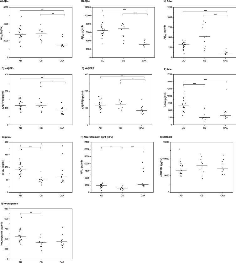
Results: We included 10 CAA, 20 AD, and 10 CS participants (mean age 68.6, 62.5, and 62.2 years, respectively). In unadjusted analyses, CAA patients had a distinctive CSF biomarker profile, with significantly lower (p < 0.01) median concentrations of Aβ38, Aβ40, Aβ42, sAβPPα, and sAβPPβ. CAA patients had higher levels of neurofilament light (NFL) than the CS group (p < 0.01), but there were no significant differences in CSF total tau, phospho-tau, soluble TREM2 (sTREM2), or neurogranin concentrations. AD patients had higher total tau, phospho-tau and neurogranin than CS and CAA groups. In age-adjusted analyses, differences for the CAA group remained for Aβ38, Aβ40, Aβ42, and sAβPPβ. Comparing CAA patients with amyloid-PET positive (n = 5) and negative (n = 5) scans, PET positive individuals had lower (p < 0.05) concentrations of CSF Aβ42, and higher total tau, phospho-tau, NFL, and neurogranin concentrations, consistent with an "AD-like" profile.
Conclusion: CAA has a characteristic biomarker profile, suggestive of a global, rather than selective, accumulation of amyloid species; we also provide evidence of different phenotypes according to amyloid-PET positivity. Further replication and validation of these preliminary findings in larger cohorts is needed.
Keywords: Alzheimer’s disease; amyloid-β; biomarkers; cerebral amyloid angiopathy; cerebrospinal fluid.
Conflict of interest statement
Authors’ disclosures available online (
Figures
References
-
- Knudsen KA, Rosand J, Karluk D, Greenberg SM (2001) Clinical diagnosis of cerebral amyloid angiopathy: Validation of the Boston criteria. Neurology 56, 537–539. - PubMed
-
- Banerjee G, Werring DJ (2019) Feasibility of clinical trial recruitment for cerebral amyloid angiopathy: A specialist single centre experience. J Neurol Sci 409, 116580. - PubMed
-
- Weston PS, Paterson RW, Modat M, Burgos N, Cardoso MJ, Magdalinou N, Lehmann M, Dickson JC, Barnes A, Bomanji JB, Kayani I, Cash DM, Ourselin S, Toombs J, Lunn MP, Mummery CJ, Warren JD, Rossor MN, Fox NC, Zetterberg H, Schott JM (2015) Using florbetapir positron emission tomography to explore cerebrospinal fluid cut points and gray zones in small sample sizes. Alzheimers Dement (Amst) 1, 440–446. - PMC - PubMed
Publication types
MeSH terms
Substances
Grants and funding
LinkOut - more resources
Full Text Sources
Medical
