Review
. 2020;74(1):1-28.
doi: 10.3233/JAD-191150.

Inflammation in Traumatic Brain Injury

Affiliations
Review

Inflammation in Traumatic Brain Injury

Teodor T Postolache et al. J Alzheimers Dis. 2020.

Abstract

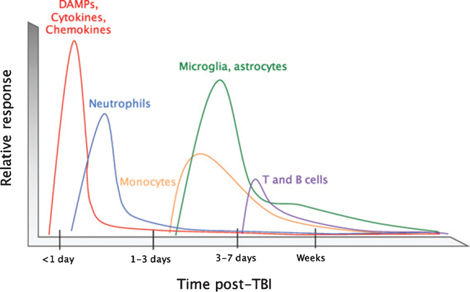
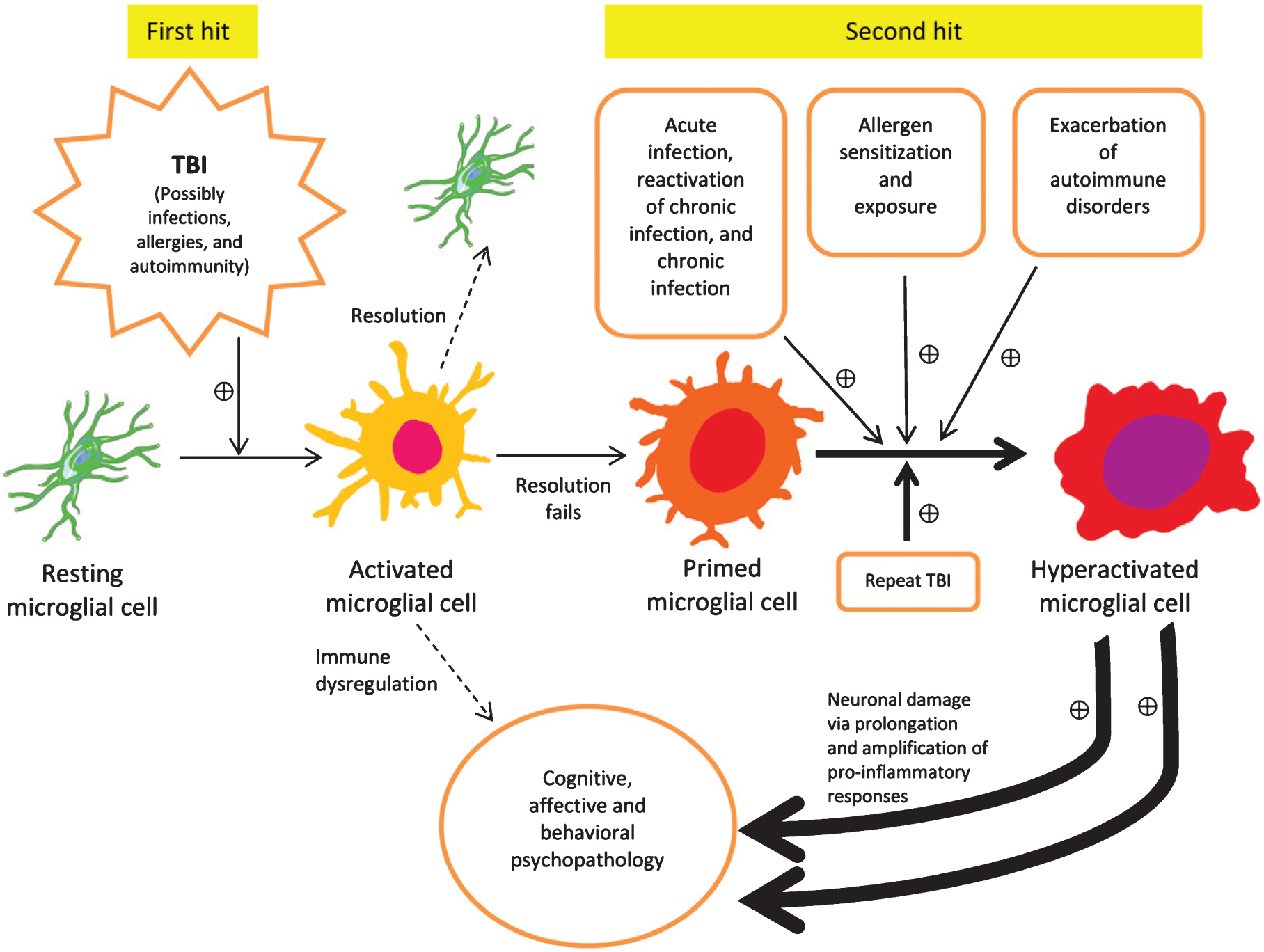
There is an increasing evidence that inflammation contributes to clinical and functional outcomes in traumatic brain injury (TBI). Many successful target-engaging, lesion-reducing, symptom-alleviating, and function-improving interventions in animal models of TBI have failed to show efficacy in clinical trials. Timing and immunological context are paramount for the direction, quality, and intensity of immune responses to TBI and the resulting neuroanatomical, clinical, and functional course. We present components of the immune system implicated in TBI, potential immune targets, and target-engaging interventions. The main objective of our article is to point toward modifiable molecular and cellular mechanisms that may modify the outcomes in TBI, and contribute to increasing the translational value of interventions that have been identified in animal models of TBI.

Keywords: Depression; glia; immune challenge; immunomodulation; inflammation; priming; probiotic; traumatic brain injury.


Figures

Fig. 1.
Fig. 1.
Timeline of the immune response to TBI. After impact, rapid tissue release of damage-associated molecular patterns (DAMPs) prompts resident cells to secrete chemokines and cytokines. These molecules attract neutrophils, which contribute to circumscribing the injury site and promoting the removal of damaged and dead cells and debris. Infiltrating monocytes and activated glia begin to predominate 3–5 days post-injury to defend against infection and perform reparative functions. T and B cells can also be recruited to sites of brain pathology at later time points in the response (3–7 days post-injury). The timeline is modeled to human TBI across modalities, and is likely to correspond to the great majority of animal models of TBI (Reproduced from McKee CA, Lukens JR (2016) Emerging roles for the immune system in traumatic brain injury. Front Immunol 7, 556).
Fig. 2.
Fig. 2.
Traumatic brain injury-induced macrophage response varies in reaction to immune stressors that occur before, with, and after the injury. A solid black line and gray shading depicts normal, age-related health burden. A) Traumatic brain injury (TBI) occurring in the absence of Aβ (dotted black line) or tau results in acute macrophage-related neuroinflammation that subsides over time. TBI in the presence of Aβ (solid red line) results in an acute blunted macrophage response that increases at chronic post-injury time points. TBI in the presence of pathological tau (solid blue line) results in an enhanced macrophage response to TBI that remains elevated at chronic post-injury time points. B) Over time, macrophage-related neuroinflammation increases with normal health burden. Single TBI (dotted black line) results in acute macrophage-related neuroinflammation that subsides over time. Pre-injury peripheral immune challenge at sub-threshold levels (red line) attenuates the post-injury macrophage-related inflammatory response to TBI. Post-injury peripheral immune challenge (solid blue line) causes a hyper-active macrophage response correlating with behavioral dysfunction. Repetitive post-injury immune challenge (dotted blue line), similar to what is observed in repetitive TBI, increases macrophage-related neuroinflammation and correlates with the advanced neuropathology. The figure represents a model of what occurs in human TBI, translating specific findings in rodent TBI studies. (Reproduced from Kokiko-Cochran ON, Godbout JP (2018) The inflammatory continuum of traumatic brain injury and Alzheimer’s disease. Front Immunol 9, 672.)
Fig. 3.
Fig. 3.
Double hit model illustrates how during the first hit microglia become activated, and then potentially primed. A second hit may include any source of inflammation, such as intense stress, autoimmune disease reactivation, allergen exposure in atopic individuals, and most commonly, infections (acute, chronic, chronic reactivated) and/or repeat TBI. The primed microglia convey a risk for developing behavioral, affective, and cognitive disorders, and exposure to subsequent inflammation driven “hits” exponentially increases that probability, as well as symptomatic intensity and functional deterioration. The double hit model is expected to fully represent all TBI models in rodents, as well as its translation to clinical TBI, with the difference that in human TBI, a multitude of individual immune stressors interact with TBI in contrast to experimental animals living in conditions characterized by limited variation between individuals in exposure to various immune modifiers. This is a top-of-the-list possible explanation of why immune targeting interventions for TBI that are beneficial in rodents do not translate in humans, with few exceptions (e.g., aerobic exercise).

References

    1. Fehily B, Fitzgerald M (2017) Repeated mild traumatic brain injury: potential mechanisms of damage. Cell Transplant 26, 1131–1155. - PMC - PubMed
    1. Lozano D, Gonzales-Portillo GS, Acosta S, de la Pena I, Tajiri N, Kaneko Y, Borlongan CV (2015) Neuroinflammatory responses to traumatic brain injury: etiology, clinical consequences, and therapeutic opportunities. Neuropsychiatr Dis Treat 11, 97–106. - PMC - PubMed
    1. Silver JM, McAllister TW, Arciniegas DB (2019) Textbook of traumatic brain injury. American Psychiatric Publishing.
    1. Burda JE, Sofroniew MV (2014) Reactive gliosis and the multicellular response to CNS damage and disease. Neuron 81, 229–248. - PMC - PubMed
    1. Moalem G, Leibowitz-Amit R, Yoles E, Mor F, Cohen IR, Schwartz M (1999) Autoimmune T cells protect neurons from secondary degeneration after central nervous system axotomy. Nat Med 5, 49–55. - PubMed

Publication types