Review
. 2020 Jun;160(2):157-170.
doi: 10.1111/imm.13193. Epub 2020 Apr 15.

The liver as an immunological barrier redefined by single-cell analysis

Affiliations
Review

The liver as an immunological barrier redefined by single-cell analysis

Zania Stamataki et al. Immunology. 2020 Jun.

Abstract

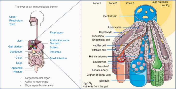
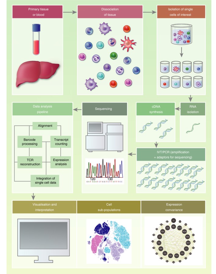
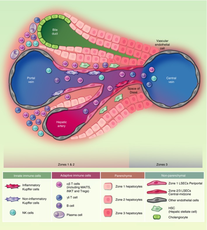
The liver is a front-line immune tissue that plays a major role in the detection, capture and clearance of pathogens and foreign antigens entering the bloodstream, especially from the gut. Our largest internal organ maintains this immune barrier in the face of constant exposure to external but harmless antigens through a highly specialized network of liver-adapted immune cells. Mapping the immune resident compartment in the liver has been challenging because it requires multimodal single-cell deep phenotyping approaches of often rare cell populations in difficult to access samples. We can now measure the RNA transcripts present in a single cell (scRNA-seq), which is revolutionizing the way we characterize cell types. scRNA-seq has been applied to the diverse array of immune cells present in murine and human livers in health and disease. Here, we summarize how emerging single-cell technologies have advanced or redefined our understanding of the immunological barrier provided by the liver.

Keywords: RNA-seq; immune barrier; liver; liver resident cells; single cells; transcriptomics.


Conflict of interest statement

The authors declare no conflict of interest.

Figures

Figure 1
Figure 1
The liver as an immunological barrier. The liver receives a dual blood supply of oxygen‐rich arterial blood from the heart and nutrient/antigen‐rich venous blood, that brings leucocytes that have passed through the gastrointestinal tract, pancreas and spleen. Immune cells from the hepatic artery and portal vein mix in the liver sinusoids and drain into the central vein. The differential access to oxygen and nutrients proximal to the portal triad (hepatic artery, portal vein, bile duct) compared with the oxygen‐ and nutrient‐poor central vein regions affects hepatocyte morphology and forms the basis for defining liver zonation. O2, partial pressure of oxygen.
Figure 2
Figure 2
Single‐cell RNA sequencing (scRNA‐seq) experimental workflow. The standard workflow for scRNA‐seq (reviewed in detail in Refs 14, 15) involves: isolation of single cells (commonly by flow cytometric sorting or microfluidics), RNA extraction, enrichment of mRNA [olig‐dT‐enrichment of poly(A) tail mRNA] or depletion of ribosomal RNA (accounting for up to 95% of cellular RNA) before reverse transcription to complementary DNA (cDNA). cDNA is amplified, sequencing adaptors added, before fragmentation to produce a library of short cDNA molecules for sequencing. The resulting sequences are aligned to a reference genome, and the relative quantification of mRNA molecules is calculated for each gene per cell. Statistical modelling is then employed to identify significant differences in the expression level of genes (differential gene expression analysis) between groups of cells and/or samples. cDNA, complementary DNA; PCR, polymerase chain reaction; TCR, T‐cell receptor; IVT, in vitro transcription. Example covariance map created using Genemania. 129
Figure 3
Figure 3
Human liver cell types. Experimental data from single‐cell analyses have provided a broad list of the cell subtypes that are present in the healthy human liver. This schematic shows the location of these parenchymal and non‐parenchymal cell subsets across the portal‐venous sinusoidal axis according to immunohistochemistry and spatial cell sorting where available. Many immune subsets have only been investigated in cell suspension from liver samples without confirmatory spatial staining, as such their locations across this axis are not representative (i.e. αβ‐T‐cells, γδ‐T‐cells, NK cells, B‐cells and plasma cells). Endothelial cells are represented as periportal zone 1 liver sinusoidal endothelial cells (LSECs) and zone 2/3 central‐midzone LSECs, 23 , 40 , 53 and non‐LSEC endothelial cells (representing endothelial cells around the portal vein, central vein, hepatic artery). ‘Inflammatory’ central‐venous Kupffer cells and ‘non‐inflammatory’ periportal Kupffer cells are represented. 23 , 40 , 62 It is important to consider cell−cell interactions between liver‐adapted and liver‐infiltrating cells and hepatocytes. A complex network of cell−cell interactions inform the liver immune barrier, for example LSEC−T‐cell interaction can lead to T‐cell anergy if T‐cells recognize antigen in the absence of co‐stimulation, 28 and T‐cell−hepatocyte interactions may skew T‐cell responses towards a regulatory phenotype (for review, see Ref. 130). Hepatocytes respond to inflammation by producing immunomodulatory cytokines, such as IL‐6, 30 , 34 and can even engulf immune cells, for instance enclysis of CD4+ T‐cells, 51 to influence immune responses in the liver. HSCs, hepatocyte, endothelial cell‐derived signals combine to drive Kupffer cell differentiation; 60 αβ‐T‐cell, alpha‐beta T‐cell receptor expressing T‐cell; γδ‐T‐cell, gamma‐delta T‐cell receptor expressing T‐cell; LSEC, liver sinusoidal endothelial cells; MAITs, mucosal‐associated invariant T‐cells; NK, natural killer; iNKT, invariant natural killer cell; Treg, regulatory T‐cell.

References

    1. Paulos CM, Wrzesinski C, Kaiser A, Hinrichs CS, Chieppa M, Cassard L et al Microbial translocation augments the function of adoptively transferred self/tumor‐specific CD8+ T cells via TLR4 signaling. J Clin Invest 2007; 117:2197–204. - PMC - PubMed
    1. Kubes P, Jenne C. Immune responses in the liver. Annu Rev Immunol 2018; 36:247–77. - PubMed
    1. Son G, Kremer M, Hines IN. Contribution of gut bacteria to liver pathobiology. Gastroenterol Res Pract 2010; 2010:453 563. - PMC - PubMed
    1. Huang H, Lu Y, Zhou T, Gu G, Xia Q. Innate immune cells in immune tolerance after liver transplantation. Front Immunol 2018; 9:2401. - PMC - PubMed
    1. Crispe IN. Immune tolerance in liver disease. Hepatology 2014; 60(6):2109–17. - PMC - PubMed

Publication types