. 2020 Aug;35(8):1572-1581.
doi: 10.1002/jbmr.4009. Epub 2020 Apr 2.

Intervertebral Disc Degeneration Is Associated With Aberrant Endplate Remodeling and Reduced Small Molecule Transport

Affiliations

Intervertebral Disc Degeneration Is Associated With Aberrant Endplate Remodeling and Reduced Small Molecule Transport

Beth G Ashinsky et al. J Bone Miner Res. 2020 Aug.

Abstract

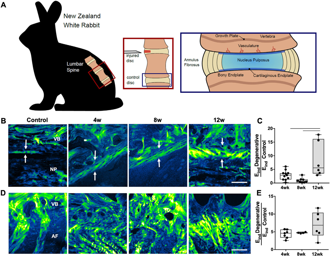
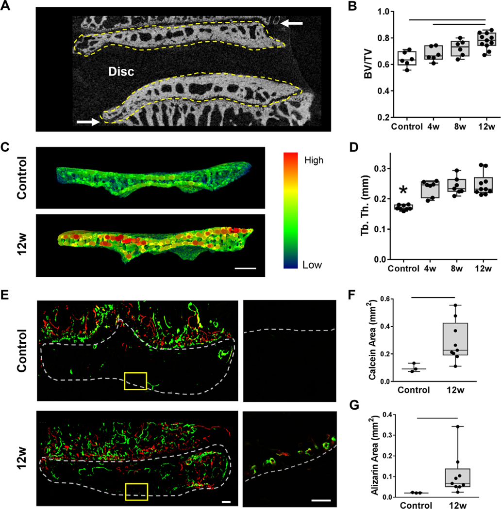
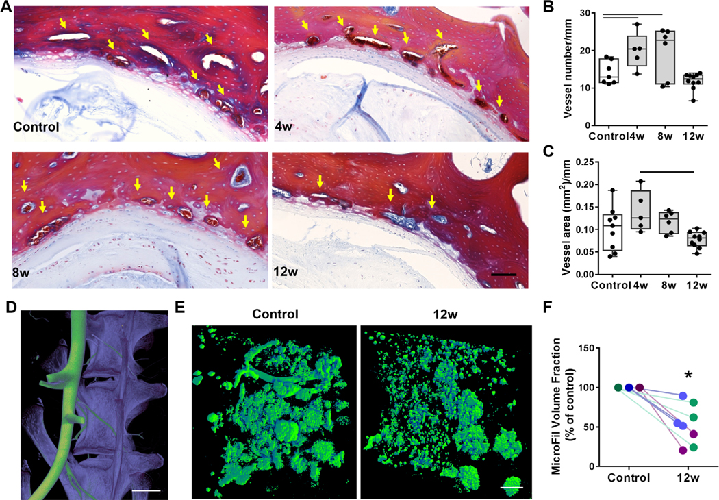
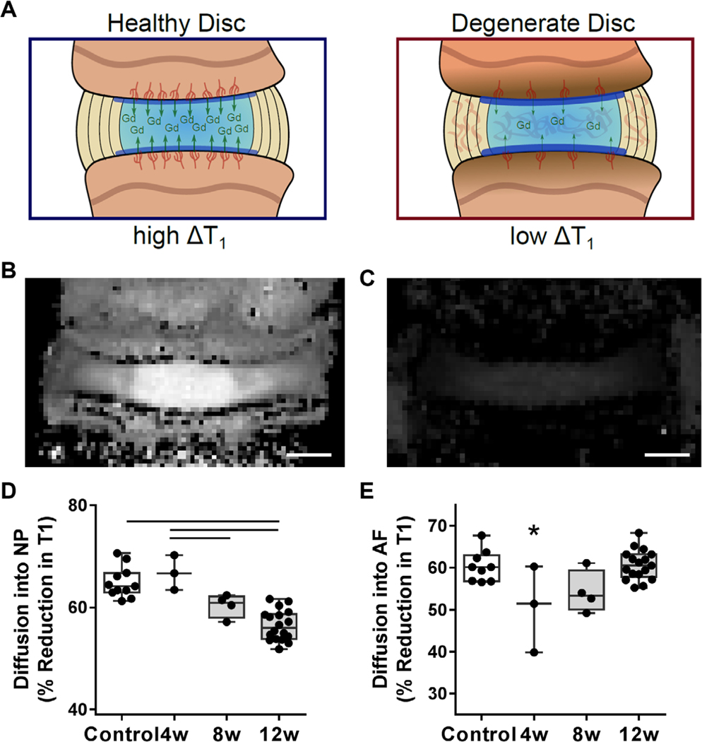
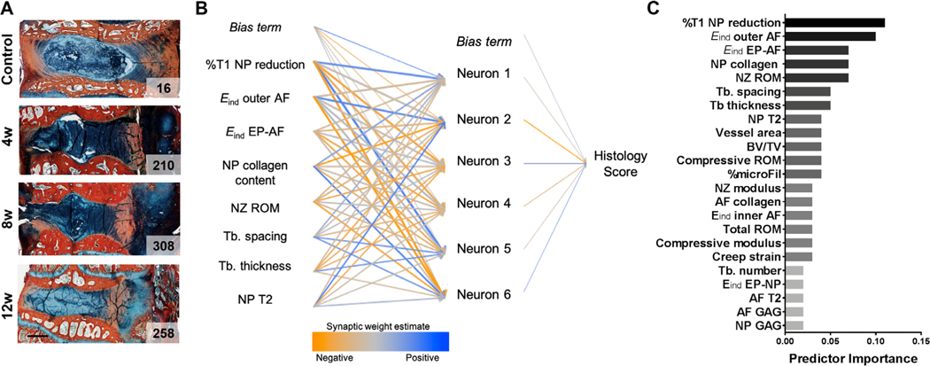
The intervertebral disc is the largest avascular structure in the body, and cells within the disc rely on diffusive transport via vasculature located within the vertebral endplate to receive nutrients, eliminate waste products, and maintain disc health. However, the mechanisms by which small molecule transport into the disc occurs in vivo and how these parameters change with disc degeneration remain understudied. Here, we utilize an in vivo rabbit puncture disc degeneration model to study these interactions and provide evidence that remodeling of the endplate adjacent to the disc occurs concomitant with degeneration. Our results identify significant increases in endplate bone volume fraction, increases in microscale stiffness of the soft tissue interfaces between the disc and vertebral bone, and reductions in endplate vascularity and small molecule transport into the disc as a function of degenerative state. A neural network model identified changes in diffusion into the disc as the most significant predictor of disc degeneration. These findings support the critical role of trans-endplate transport in disease progression and will improve patient selection to direct appropriate surgical intervention and inform new therapeutic approaches to improve disc health. © 2020 American Society for Bone and Mineral Research. Published 2020. This article is a U.S. Government work and is in the public domain in the USA.

Keywords: ANIMAL MODELS; BONE REMODELING; INTERVERTEBRAL DISC DEGENERATION; SMALL MOLECULE DIFFUSION; VASCULARITY.


Conflict of interest statement

Disclosures

RLM is the editor of JOR Spine. All other authors state that they have no conflicts of interests.

Figures

Fig. 1.
Fig. 1.
(A) Schematic depicting the surgical approach for the disc puncture model and regions of the spinal motion segment investigated in this study. (B) Second harmonic generation imaging (SHG) of the cartilaginous endplate between the vertebral endplate and nucleus pulposus and (C) corresponding indentation modulus (Eind) of this region, normalized to control values. (D) SHG of the vertebral endplate-AF region and corresponding Eind of this region, normalized to control values. Arrows indicate cartilage endplate thickness. VB = vertebral body; NP = nucleus pulposus; AF = annulus fibrosus. Scale bar = 100 μm. Bars denote statistical significance (4 to 12 weeks, p = 0.008; 8 to 12 weeks, p = 0.001), Kruskal-Wallis test.
Fig. 2.
Fig. 2.
(A) Representative mid-coronal μCT slice of a control motion segment, with the cranial and caudal vertebral endplates outlined in yellow, denoting the volume of interest analyzed for bone morphometry. Arrows indicate the growth plates. (B) Quantification of bone volume/total volume (BV/TV) in the endplate regions for each group. (C) Representative trabecular thickness (Tb.Th) map of the endplate volume of interest and (D) corresponding quantification of Tb.Th of the endplates in each group. (E) Representative images of the bone fluorochrome labels (calcein = green, alizarin = red) in the endplate region. The dashed outline in the macroscale image denotes the endplate region between the disc and growth plate (scale bar = 100 μm). In the inset images (scale bar = 100 μm), the dashed line denotes the border between the NP and the endplate. (F) Quantification of calcein and alizarin areas in the control and 12-week punctured groups. Bars denote statistical significance (BV/TV = control-12 weeks, p = 0.0004; 4–12 weeks, p = 0.01; Tb.Th = control-4 weeks, p = 0.0006; control-8 weeks, p = 0.0002; control-12 weeks, p < 0.0001, ANOVA). Calcein: p = 0.01; alizarin: p = 0.009, Mann–Whitney test.
Fig. 3.
Fig. 3.
(A) Mallory-Heidenhain staining of histology sections for the endplate vasculature; arrows indicate vessels. Scale bar = 100 μm. (B) Quantification of vessel number and (C) area normalized to endplate length adjacent to the NP. 3D μCT reconstructions of the (D) spinal vasculature perfused with microFil (green) and the vertebral bodies (purple) before decalcification (scale bar = 5 mm) and (E) microFil perfusion of the small vasculature adjacent to the disc, visualized after decalcification (scale bar = 500 μm). (F) MicroFil volume fraction in the endplate adjacent to degenerative discs 12 weeks post-puncture compared with controls (each color indicates separate animal). Bars denote statistical significance (vessel area = control-4 weeks, p = 0.01; 4–8 weeks, p = 0.009; vessel number = p = 0.03), Kruskal-Wallis test.
Fig. 4.
Fig. 4.
(A) Schematic depicting the change in T1 relaxation time within the intervertebral disc after administration of the contrast agent gadodiamide (Gd). Representative MRI T1 maps of a control disc (B) before and (C) after administration of Gd, demonstrating the reduction in T1 relaxation time (scale bar = 1 mm). (D) The percent reduction in T1 into the NP and (E) AF after Gd administration in each experimental group. Bars denote statistical significance (%T1 NP = control-12 weeks, p < 0.0001; 4–8 weeks, p = 0.03; 4–12 weeks, p < 0.0001; %T1 AF = control-4 weeks, p = 0.01; 4–12 weeks, p = 0.006), ANOVA.
Fig. 5.
Fig. 5.
(A) Representative Alcian blue and picrosirius red stained sections from each experimental group, with total histology score indicated on each image (scale bar = 2 mm). (B) A neural network model was utilized to predict histology score based on 23 input parameters of disc structure and function (the top eight predictors are shown). (C) Relative predictor importance for all input parameters.

Similar articles

Cited by

References

    1. Cassidy JJ, Hiltner A, Baer E. Hierarchical structure of the intervertebral disc. Connect Tissue Res. 1989;23(1):75–88. - PubMed
    1. Korecki CL, MacLean JJ, Iatridis JC. Dynamic compression effects on intervertebral disc mechanics and biology. Spine. 2008;33(13): 1403–9. - PMC - PubMed
    1. Fields AJ, Ballatori A, Liebenberg EC, Lotz JC. Contribution of the end-plates to disc degeneration. Curr Mol Biol Rep. 2018;4(4):151–60. - PMC - PubMed
    1. Huang Y-sC, Urban JPG, Luk KDK. Intervertebral disc regeneration: do nutrients lead the way? Nat Rev Rheumatol. 2014;10(9):561–6. - PubMed
    1. Roberts S, Menage J, Urban JPG. Biochemical and structural properties of the cartilage end-plate and its relation to the intervertebral disc. Spine. 1989;14(2):166–74. - PubMed

Publication types

MeSH terms