. 2020 Mar 12;21(6):1951.
doi: 10.3390/ijms21061951.

Humanization of Tumor Stroma by Tissue Engineering as a Tool to Improve Squamous Cell Carcinoma Xenograft

Affiliations

Humanization of Tumor Stroma by Tissue Engineering as a Tool to Improve Squamous Cell Carcinoma Xenograft

Sara Guerrero-Aspizua et al. Int J Mol Sci. .

Abstract

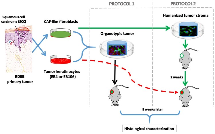
The role of stroma is fundamental in the development and behavior of epithelial tumors. In this regard, limited growth of squamous cell carcinomas (SCC) or cell-lines derived from them has been achieved in immunodeficient mice. Moreover, lack of faithful recapitulation of the original human neoplasia complexity is often observed in xenografted tumors. Here, we used tissue engineering techniques to recreate a humanized tumor stroma for SCCs grafted in host mice, by combining CAF (cancer associated fibroblasts)-like cells with a biocompatible scaffold. The stroma was either co-injected with epithelial cell lines derived from aggressive SCC or implanted 15 days before the injection of the tumoral cells, to allow its vascularization and maturation. None of the mice injected with the cell lines without stroma were able to develop a SCC. In contrast, tumors were able to grow when SCC cells were injected into previously established humanized stroma. Histologically, all of the regenerated tumors were moderately differentiated SCC with a well-developed stroma, resembling that found in the original human neoplasm. Persistence of human stromal cells was also confirmed by immunohistochemistry. In summary, we provide a proof of concept that humanized tumor stroma, generated by tissue engineering, can facilitate the development of epithelial tumors in immunodeficient mice.

Keywords: CAF; SCC; stroma; tissue engineering; tumor; xenografts.


Conflict of interest statement

The authors declare no conflict of interest.

Figures

Figure 1
Figure 1
Tissue engineering humanized stroma for development of squamous cell carcinomas (SCC) in immunodeficient mice: two different approaches were followed. In protocol 1 simultaneous injection of tumoral cells and stroma (cancer associated fibroblasts (CAF)-like cells and biocompatible matrix) was performed, whereas in protocol 2 the generation of a humanized stroma was performed 2 weeks before the injection of the neoplastic keratinocytes. SCC-cells and stromal fibroblasts derived from recessive dystrophic epidermolysis bullosa (RDEB) patients were used during this experimentation, given their high predisposition of RDEB patients to develop very aggressive SCC. Moreover, the similarities of the molecular profile between RDEB dermal cells with that of CAFs, which promotes the invasion of tumor cells, has been widely studied [6,13,14,15,16,17]. In the first approach, to assess the tumorigenic potential of the SCC-derived cell lines used, immunodeficient mice were injected with tumor keratinocytes (EB4 or EB106), without the addition of exogenous stroma (2 × 10⁶ cells/animal. n = 3 in each experimental group). No tumor was detected after 60 days post-injection.
Figure 2
Figure 2
Macroscopic characterization of tumors developed in the presence of humanized stroma after 8 weeks of transplantation. (a) Tumor developed by protocol 1; (b) tumor developed by protocol2.
Figure 3
Figure 3
Persistence of tumor stromal cells in bioengineered SCC following protocols 1 and 2, respectively. (a) In vitro fluorescence microscope image from the cell culture, before transplantation, where both the EGFP-CAFs and RFP-SCC-keratinocytes are observed; scale bar: 200 μm. (b) In vivo3D fluorescence image obtained from the tumor, 8 weeks after implantation, using XLView Loupe. Scale bar: 2 mm.
Figure 4
Figure 4
Histopathological analysis of representative tumors generated by different protocols of SCC stroma humanization. (a–d) Protocol 1; (e–h) protocol 2; (a) and (e)hematoxylin-eosin staining shows the structure of the tumors developed, as evidenced by the presence of tumor nests and cornified pearls and blood vessels in protocol 2; (b) and (f) Mason trichrome staining shows the different humanized tumoral stroma in both protocols;(c) and (g) show the human origin of fibroblasts surrounding the tumor structure by a h-vimentin staining; (d) and (h)show that tumors developed by protocol 2 have more proliferative cells as shown by a Ki67 staining.(i) Shows a quantification of the epithelial component of the tumor where ki67+ nuclei/100 epithelial cells is shown. For this calculation a total number of ten representative fields were included. Images are representative results for each group (n = 3). Scale bars 200 μm.

Similar articles

Cited by

References

    1. Bremnes R.M., Donnem T., Al-Saad S., Al-Shibli K., Andersen S., Sirera R., Camps C., Marinez I., Busund L.T. The role of tumor stroma in cancer progression and prognosis: Emphasis on carcinoma-associated fibroblasts and non-small cell lung cancer. J. Thorac. Oncol. 2011;6:209–217. doi: 10.1097/JTO.0b013e3181f8a1bd. - DOI - PubMed
    1. De Wever O., Mareel M. Role of tissue stroma in cancer cell invasion. J. Pathol. 2003;200:429–447. doi: 10.1002/path.1398. - DOI - PubMed
    1. Chan T.S., Shaked Y., Tsai K.K. Targeting the Interplay Between Cancer Fibroblasts, Mesenchymal Stem Cells, and Cancer Stem Cells in Desmoplastic Cancers. Front. Oncol. 2019;9:688. doi: 10.3389/fonc.2019.00688. - DOI - PMC - PubMed
    1. Lindner T., Loktev A., Giesel F., Kratochwil C., Altmann A., Haberkorn U. Targeting of activated fibroblasts for imaging and therapy. EJNMMI Radiopharm. Chem. 2019;4:16. doi: 10.1186/s41181-019-0069-0. - DOI - PMC - PubMed
    1. Ao M., Franco O.E., Park D., Raman D., Williams K., Hayward S.W. Cross-talk between paracrine-acting cytokine and chemokine pathways promotes malignancy in benign human prostatic epithelium. Cancer Res. 2007;67:4244–4253. doi: 10.1158/0008-5472.CAN-06-3946. - DOI - PubMed