. 2020 Mar 12;12(3):661.
doi: 10.3390/cancers12030661.

Characterization of Human NK Cell-Derived Exosomes: Role of DNAM1 Receptor In Exosome-Mediated Cytotoxicity Against Tumor

Affiliations

Characterization of Human NK Cell-Derived Exosomes: Role of DNAM1 Receptor In Exosome-Mediated Cytotoxicity Against Tumor

Anna Laura Di Pace et al. Cancers (Basel). .

Abstract

Despite the pivotal role of natural killer (NK) cells in defenses against tumors, their exploitation in cancer treatment is still limited due to their reduced ability to reaching tumor sites and the inhibitory effects of tumor microenvironment (TME) on their function. In this study, we have characterized the exosomes from IL2- or IL15-cultured human NK cells. Both cytokines induced comparable amounts of exosomes with similar cargo composition. Analysis of molecules contained within or exposed at the exosome surface, allowed the identification of molecules playing important roles in the NK cell function including IFN-γ, Lymphocyte Function-Associated Antigen (LFA-1), DNAX Accessory Molecule-1 (DNAM1) and Programmed Cell Death Protein (PD-1). Importantly, we show that DNAM1 is involved in exosome-mediated cytotoxicity as revealed by experiments using blocking antibodies to DNAM1 or DNAM1 ligands. In addition, antibody-mediated inhibition of exosome cytotoxicity results in a delay in target cell apoptosis. We also provide evidence that NK-exosomes may exert their cytolytic activity after short time interval and even at low concentrations. Regarding their possible use in immunotherapy, NK exosomes, detectable in peripheral blood, can diffuse into tissues and exert their cytolytic effect at tumor sites. This property offers a clue to integrate cancer treatments with NK exosomes.

Keywords: DNAM1; NK cells; cancer; cytotoxicity; exosomes; tumor therapy.


Conflict of interest statement

The authors declare no conflict of interest

Figures

Figure 1
Figure 1
Characterization of exosomes released by human natural killer (NK) cells. (A) Flow chart for NK cell culture and NK exosome isolation. (B) Nano-particle size distribution curve and picture of NK-derived exosomes by nanotracking analysis (NTA). One out of 4 representative preparation of exosomes is shown. Mean and mode numbers are reported. Bar: 500 nm (C) Flow-cytometry analysis of CD81 and CD63 expression on beads-conjugated exosomes derived from NK cells. Filled grey profiles and black lines represent control isotype and samples stained with CD81 or CD63, respectively. (D) Western blot analysis of CD81 (23 kDa), CD63 (30-60 kDa), TSG101 (43 kDa), and Calnexin (90 kDa) expression on NK cell lysates compared to NK-derived exosomes.
Figure 2
Figure 2
Phenotypic analysis of NK-derived exosomes. (A) Mean ± SD (standard deviation) of total amount (μg of exosomes/106 cells) of secreted exosomes by IL2- and IL15-stimulated NK cells after 48 h of culture (n = 3). (BD) Flow-cytometry analysis of indicated markers (black lines) on exosomes isolated from IL2- and IL15-stimulated NK cells. Filled grey profiles represent controls. One representative experiment out of 3 performed is shown. (B) Evaluation of surface antigens in NK-derived exosomes. (C) Analysis of cytotoxic proteins present inside NK-derived exosomes by flow-cytometry. (D) Expression of novel surface and inner markers in exosomes from IL2-stimulated NK cells by flow-cytometry (LFA-1, DNAM1, IFN-γ and PD1).
Figure 3
Figure 3
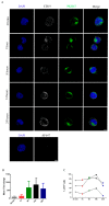
Uptake of PKH67+ NK-derived exosomes to NALM-18 lymphoma cell line. (A) Confocal microscopy analysis of exosome internalization by NALM-18 target cells at different time points (30 min, 1 h, 8 h, 14 h, and 24 h). Cells, stained with anti-CD19 antibody (white) and DAPI (blue), were incubated with 20 µg of PKH67-labelled NK exosomes (green) and their internalization was evaluated at different times (Upper figure). Bar: 5 μm. NALM-18 cells, stained with DAPI (blue) and incubated with Alexa Fluor 647 conjugated secondary antibody as control for antibody specificity. (Lower figure) Bar: 5 μm. (B) Exosome uptake evaluation by flow-cytometry. Fluorescence intensities of PKH+ NALM-18 cells are shown as mean fold change (n = 3). (C) Percentage of PI+ NALM-18 cells treated at different time points (30 min, 1 h, 8 h, 14 h, and 24 h) using 20 µg of NK-exosomes (n = 3).
Figure 4
Figure 4
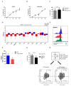
Cytotoxicity and molecular mechanisms involved in killing of K562 and NALM-18 tumor cell lines by NK-derived exosomes. (A,B) Cytotoxic assay on K562 (A) and NALM-18 (B) target cells incubated with different doses of NK-derived exosomes (1γ, 5γ, 10γ, 20γ, and 50γ). The percentage of PI+ cells was evaluated after 24 h of incubation with NK-exosomes. Mean ± SEM (Standard error of mean) of four independent experiments is shown. (C) Cytotoxic assay on NALM-18 cells treated for 24 h with 20γ of NK-derived exosomes in the absence (grey bars) or in the presence (black bars) of blocking antibodies to LFA1 ligand (CD54) (n = 4). Mean ± SEM is shown (ns = not significant). (D) Gene expression analysis in different tumors (red boxes) and healthy controls (blue boxes). TGCA datasets have been used for this analysis. For tumor abbreviations, see Table S1. (E) Analysis of DNAM1 in lung tumor biopsy and metastatic lesions by flow-cytometry. DNAM1 expression on infiltrating NK cell of primary tumor (red profile) and derived metastatic lesion (green profile) compared to those of surrounding normal tissue. One representative experiment is shown. (F) Analysis of DNAM1 expression in NK cells present in peripheral blood of lung cancer patients (n = 3) as compared to peripheral blood mononuclear cells (PBMC) from healthy donors (n = 4) by flow-cytometry. Mean fold change ± SEM is shown. (G) Cytotoxic assay on NALM-18 cells incubated with 20γ of NK exosomes in absence (grey bars) or presence (black bar) of DNAM1 ligands (CD155+CD112) blocking antibodies (n = 6) (* = p-value < 0.05). (H) Detection of active caspase in NALM-18 cells incubated with NK exosomes in the absence or in the presence of DNAM1-ligands blocking antibodies (n = 3).

References

    1. Vivier E., Tomasello E., Baratin M., Walzer T., Ugolini S. Functions of natural killer cells. Nat. Immunol. 2008;9:503–510. doi: 10.1038/ni1582. - DOI - PubMed
    1. Moretta L., Ciccone E., Mingari M.C., Biassoni R., Moretta A. Human natural killer cells: Origin, clonality, specificity, and receptors. Adv. Immunol. 1994;55:341–380. - PubMed
    1. Martin-Fontecha A., Thomsen L.L., Brett S., Gerard C., Lipp M., Lanzavecchia A., Sallusto F. Induced recruitment of NK cells to lymph nodes provides IFN-gamma for T(H)1 priming. Nat. Immunol. 2004;5:1260–1265. doi: 10.1038/ni1138. - DOI - PubMed
    1. Moretta A., Bottino C., Vitale M., Pende D., Cantoni C., Mingari M.C., Biassoni R., Moretta L. Activating receptors and coreceptors involved in human natural killer cell-mediated cytolysis. Annu. Rev. Immunol. 2001;19:197–223. doi: 10.1146/annurev.immunol.19.1.197. - DOI - PubMed
    1. Moretta L., Pietra G., Montaldo E., Vacca P., Pende D., Falco M., Del Zotto G., Locatelli F., Moretta A., Mingari M.C. Human NK cells: From surface receptors to the therapy of leukemias and solid tumors. Front. Immunol. 2014;5:87. doi: 10.3389/fimmu.2014.00087. - DOI - PMC - PubMed

LinkOut - more resources