. 2020 May:194:107999.
doi: 10.1016/j.exer.2020.107999. Epub 2020 Mar 13.

Dasatinib inhibits peripapillary scleral myofibroblast differentiation

Affiliations

Dasatinib inhibits peripapillary scleral myofibroblast differentiation

Amanda Chow et al. Exp Eye Res. 2020 May.

Abstract

Scleral fibroblast activation occurs in glaucomatous and myopic eyes. Here we perform an unbiased screen to identify kinase inhibitors that reduce fibroblast activation to diverse stimuli in vitro and to in vivo intraocular pressure (IOP) elevation. Primary cultures of peripapillary scleral (PPS) fibroblasts from two human donors were screened using a library of 80 kinase inhibitors to identify compounds that inhibit TGFβ-induced extracellular matrix (ECM) synthesis. Inhibition of myofibroblast differentiation was verified by alpha smooth muscle actin (αSMA) immunoblot and collagen contraction assay. Inhibition of IOP-induced scleral fibroblast proliferation was assessed by ELISA assay for proliferating cell nuclear antigen (PCNA). The initial screen identified 7 inhibitors as showing>80% reduction in ECM binding. Three kinase inhibitors were verified to reduce TGFβ-induced αSMA expression and cellular contractility (rottlerin, PP2, tyrphostin 9). The effect of three Src inhibitors, bosutinib, dasatinib, and SU-6656, on myofibroblast differentiation was evaluated, with only dasatinib significantly inhibiting TGFβ-induced ECM synthesis, αSMA expression, and cellular contractility at nanomolar dosages. Subconjunctival injection of dasatinib reduced IOP-induced scleral fibroblast proliferation compared to control (4.9 ± 11.1 ng/sclera with 0.1 μM versus 88.7 ± 38.6 ng/sclera in control, P < 0.0001). Dasatinib inhibits scleral myofibroblast differentiation and there is pharmacologic evidence that this inhibition is not solely due to Src-kinase inhibition.

Keywords: Fibroblast; Fibrosis; Glaucoma; Kinase inhibitor; Peripapillary sclera; Scleral remodeling; Src kinase.


Figures

Figure 1.
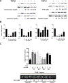
Figure 1.. PPS cell line characterization.
(a) Immunoblot for αSMA and α-tubulin (α-tub) for two primary PPS cell lines following TGFβ (1 ng/ml) treatment. (b) Densitometric analysis of (a)(n=3). (c,d) Collagen contraction without (c) and with (d) TGFβ (1 ng/ml) treatment (n=3)(cell line 1 = black bars; cell line 2 = gray bars). * P<0.05, ** P<0.01, **** P<0.0001.
Figure 2.
Figure 2.. Gelatin binding, ECM deposition assay of scleral fibroblasts.
(a) Scleral fibroblasts were treated with TGFβ for either 48 or 96 hours prior to oregon green labelled gelatin (OGG) binding and fluorescence measurement (n=4). (b) Dose response 72 hours after treatment with TGFβ (n=4). (c) Cell lines 1 (gray bars) and 2 (black bars) were treated with TGFβ for 48 hours prior to OGG absorption. * P<0.05, ** P<0.01, **** P<0.0001.
Figure 3.
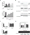
Figure 3.. Screen for kinase inhibitors that reduced TGFβ-induced OGG absorption.
linesCell lines 1 (a) and 2 (b) treated with kinase inhibitors prior to TGFβ-treatment and OGG absorption assay (n=4). Results are expressed as a ratio of OGG binding signal of kinase inhibitor treated to untreated controls (y axis) with individual compounds represented on the x-axis. (c) Overlay of (a) and (b). (d) Ven diagram showing overlap of compounds identified as strong hits in cell lines 1 and 2.
Figure 4.
Figure 4.. Kinase inhibitors reduce TGFβ-induced αSMA expression and cell contractility.
(a) Immunoblots for αSMA and α-tubulin following treatment with TGFβ and kinase inhibitors. (b) Densitometric analysis of αSMA expression relative to α-tubulin levels (n=3). (c) Collagen contraction assays using cell line 2 following treatment with TGFβ alone (control, -) and TGFβ with kinase inhibitors (n=3).
Figure 5.
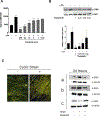
Figure 5.. Dasatinib inhibits OGG absorption αSMA expression and cell contractility.
(a) Cells were treated with bosutinib, SU-6656, or dasatinib prior to TGFβ-treatment and OGG absorption assay (n=4). (b) Immunoblot of cell lysates for αSMA and α-tubulin following treatment with TGFβ and kinase inhibitors with (c) densitometric analyses (n=3). (d) Collagen contraction assay following treatment with TGFβ alone (control, -) and TGFβ with kinase inhibitors (n=3).
Figure 6.
Figure 6.. Dasatinib prevents LPA and cyclic strain induced effects.
(a) OGG absorption assay (n=4) and (b) αSMA immunoblot with densitometry following treatment with dasatinib and LPA (10μM, n=3). (c) Immunofluorescent imaging of scleral fibroblasts (blue=DAPI stain, yellow=phalloidin (fibrillar actin)) with (+) and without (−) cyclic strain (blue line is parallel to the direction of nanotopography). αSMA immunoblot of PPS cell lines from 3 separate donors (a, b, and c) with and without dasatinib treatment (10 nM) 24 hours after exposure to cyclic strain.
Figure 7.
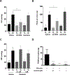
Figure 7.. Dasatinib prevents glaucoma-induced scleral proliferation.
(a) SU-6656 treatment reduces IOP and (b) PCNA levels in glaucomatous (G) eyes. (c) Dasatinib treatment did not alter IOP in control (C) or glaucomatous (G) eyes but did significantly reduce PCNA levels (d). * P<0.05, ** P<0.01, **** P<0.0001

References

    1. Voorhees AP, Jan NJ, Sigal IA. Effects of collagen microstructure and material properties on the deformation of the neural tissues of the lamina cribrosa. Acta Biomater 2017;58:278–290. - PMC - PubMed
    1. Coudrillier B, Tian J, Alexander S, Myers KM, Quigley HA, Nguyen TD. Biomechanics of the human posterior sclera: age- and glaucoma-related changes measured using inflation testing. Invest Ophthalmol Vis Sci 2012;53:1714–1728. - PMC - PubMed
    1. Norman RE, Flanagan JG, Sigal IA, Rausch SM, Tertinegg I, Ethier CR. Finite element modeling of the human sclera: influence on optic nerve head biomechanics and connections with glaucoma. Exp Eye Res 2011;93:4–12. - PubMed
    1. Hua Y, Voorhees AP, Sigal IA. Cerebrospinal Fluid Pressure: Revisiting Factors Influencing Optic Nerve Head Biomechanics. Invest Ophthalmol Vis Sci 2018;59:154–165. - PMC - PubMed
    1. Rada JA, Shelton S, Norton TT. The sclera and myopia. Exp Eye Res 2006;82:185–200. - PubMed

Publication types