. 2020 Mar 27;23(3):100958.
doi: 10.1016/j.isci.2020.100958. Epub 2020 Mar 2.

Positive Selection of a Serine Residue in Bat IRF3 Confers Enhanced Antiviral Protection

Affiliations

Positive Selection of a Serine Residue in Bat IRF3 Confers Enhanced Antiviral Protection

Arinjay Banerjee et al. iScience. .

Abstract

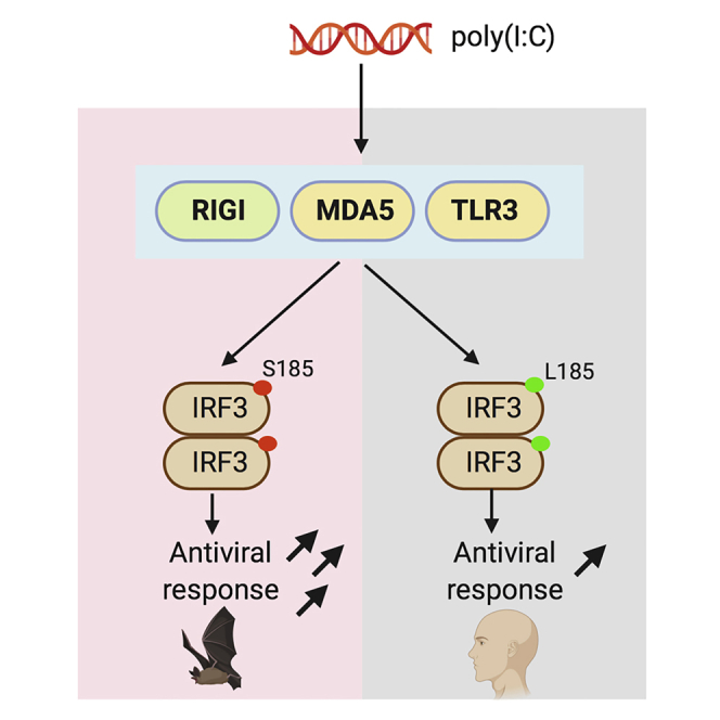
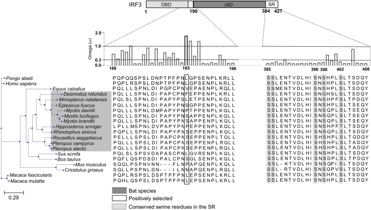
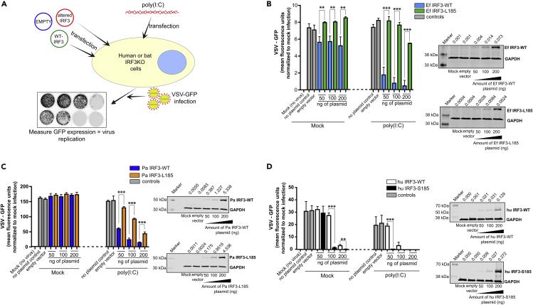
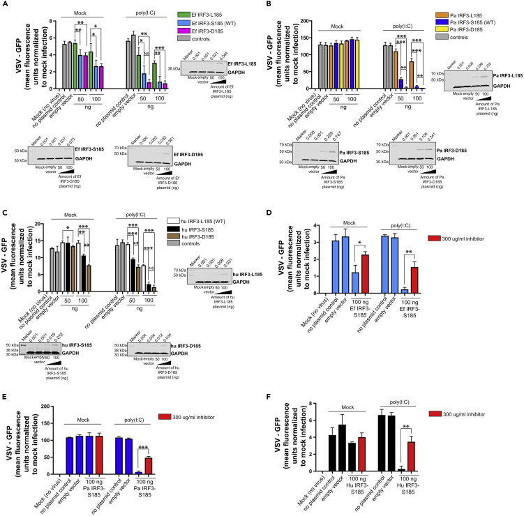
Compared with other mammals, bats harbor more zoonotic viruses per species and do not demonstrate signs of disease on infection with these viruses. To counteract infections with viruses, bats have evolved enhanced mechanisms to limit virus replication and immunopathology. However, molecular and cellular drivers of antiviral responses in bats largely remain an enigma. In this study, we demonstrate that a serine residue in IRF3 is positively selected for in multiple bat species. IRF3 is a central regulator of innate antiviral responses in mammals. Replacing the serine residue in bat IRF3 with the human leucine residue decreased antiviral protection in bat cells, whereas the addition of this serine residue in human IRF3 significantly enhanced antiviral protection in human cells. Our study provides genetic and functional evidence for enhanced IRF3-mediated antiviral responses in bats and adds support to speculations that bats have positively selected for multiple adaptations in their antiviral immune responses.

Keywords: Biological Sciences; Evolutionary Biology; Immunology.


Conflict of interest statement

Declaration of Interests The authors declare no competing interests.

Figures

None
Graphical abstract
Figure 1
Figure 1
Positive Selection of Amino Acid Residue at the 185th Position in bat IRF3 Functional domains of IRF3 are shown in the top panel above the alignment. The ratio of non-synonymous and synonymous amino acid substitutions is denoted by the omega value. Black bar indicates significant positive selection. Bat species are highlighted in dark gray. Conserved serine residues involved in human IRF3 activation are highlighted in light gray. The 185th amino acid residue in the multiple sequence alignment is highlighted by the box. DBD, DNA-binding domain; IAD, IRF association domain; SR, serine-rich region. See also Figure S1 for details on computational analysis and Table S2 for accession numbers of IRF3 sequences.
Figure 2
Figure 2
Human and Bat Cells Expressing IRF3-S185 Display Enhanced Antiviral Protection (A) Schematic representation of the experimental strategy. IRF3 knockout (KO) bat and human cells were transfected with varying concentrations of wild-type (WT) or altered IRF3 expression plasmids for 24 h. The cells were then stimulated with poly(I:C) for 6 h, followed by infection with vesicular stomatitis virus (VSV) that was engineered to express green fluorescent protein (GFP). Nineteen hours after infection, GFP expression was measured as a surrogate for virus replication. (B) VSV-GFP replication in E. fuscus IRF3 KO kidney cells (cr3-8) transfected with varying concentrations of plasmids expressing WT (S185) or altered (L185) E. fuscus IRF3 and mock treated or treated with poly(I:C) (n = 3). No plasmid and 200 ng of empty vector were used as transfection controls. Immunoblots: IRF3 protein levels in cr3-8 cells mock transfected, transfected with 200 ng empty vector (pcDNA), or transfected with varying concentrations of WT (S185) or altered (L185) IRF3 expression plasmids. (C) VSV-GFP replication in P. alecto IRF3 KO kidney cells (PakiT03-4G) transfected with varying concentrations of plasmids expressing WT (S185) or altered (L185) P. alecto IRF3 and mock treated or treated with poly(I:C) (n = 3). No plasmid and 200 ng of empty vector were used as transfection controls. Immunoblots: IRF3 protein levels in PakiT03-4G cells mock transfected, transfected with 200 ng empty vector (pcDNA) or transfected with varying concentrations of WT (S185) or altered (L185) IRF3 expression plasmids. (D) VSV-GFP replication in human IRF3 KO cells (THF-IRF3-KO) transfected with varying concentrations of plasmids expressing WT (L185) or altered (S185) human IRF3 and mock treated or treated with poly(I:C) (n = 3). No plasmid and 200 ng of empty vector were used as transfection controls. Immunoblots: IRF3 protein levels in THF IRF3 KO cells mock transfected, transfected with 200 ng empty vector (pcDNA) or transfected with varying concentrations of WT (L185) or altered (S185) IRF3 expression plasmids. Data are represented as mean ± SD, n = 3, ∗∗p < 0.01, ∗∗∗p < 0.001 (Student's t test). GFP expression is represented after normalization with mock infected cells. IRF3 protein expression and quantification data are expressed as a ratio of IRF3/GAPDH levels on top of the blots. Blots were quantified using Image Studio (LI-COR) (n = 3). KO, knockout; WT, wild-type; Ef, E. fuscus; Pa, P. alecto; Hu, human; NS, not significant.
Figure 3
Figure 3
IRF3 S185- and D185-Expressing Bat and Human Cells Mount a Robust Antiviral Response to Double-Stranded RNA (A) VSV-GFP replication in E. fuscus IRF3 KO kidney cells (cr3-8) transfected with varying concentrations of plasmids expressing L185, S185, or D185 forms of E. fuscus IRF3 and mock treated or treated with poly(I:C) (n = 3). No plasmid and 100 ng of empty vector were used as transfection controls. Immunoblots: IRF3 protein levels in cr3-8 cells mock transfected, transfected with 100 ng empty vector (pcDNA), or transfected with varying concentrations of L185, S185, and D185 IRF3 expression plasmids. (B) VSV-GFP replication in P. alecto IRF3 KO kidney cells (PakiT03-4G) transfected with varying concentrations of plasmids expressing L185, S185, or D185 forms of P. alecto IRF3 and mock treated or treated with poly(I:C) (n = 3). No plasmid and 100 ng of empty vector were used as transfection controls. Immunoblots: IRF3 protein levels in PakiT03-4G cells mock transfected, transfected with 100 ng empty vector (pcDNA), or transfected with varying concentrations of L185, S185, or D185 IRF3 expression plasmids. (C) VSV-GFP replication in human IRF3 KO cells (THF-IRF3-KO) transfected with varying concentrations of plasmids expressing L185, S185, or D185 forms of human IRF3 and mock treated or treated with poly(I:C) (n = 3). No plasmid and 100 ng of empty vector were used as transfection controls. Immunoblots: IRF3 protein levels in THF-IRF3-KO cells mock transfected, transfected with 100 ng empty vector (pcDNA), or transfected with varying concentrations of L185, S185, and D185 IRF3 expression plasmids. (D) VSV-GFP replication in E. fuscus IRF3 KO kidney cells (cr3-8) transfected with 100 ng of plasmid expressing E. fuscus IRF3-S185 and mock treated or treated with 300 μg/mL of TBK1 and IKKϵ inhibitor. After treatment with the inhibitor, cells were mock stimulated or stimulated with poly(I:C) (n = 3). Normalized VSV-GFP levels in cells treated with TBK1 and IKKϵ inhibitor are denoted by red bars. No plasmid and 100 ng of empty vector were used as transfection controls. (E) VSV-GFP replication in P. alecto IRF3 KO kidney cells (PakiT03-4G) transfected with 100 ng of plasmid expressing P. alecto IRF3-S185 and mock treated or treated with 300 μg/mL of TBK1 and IKKϵ inhibitor. After treatment with the inhibitor, cells were mock stimulated or stimulated with poly(I:C) (n = 3). Normalized VSV-GFP levels in cells treated with TBK1 and IKKϵ inhibitor are denoted by red bars. No plasmid and 100 ng of empty vector were used as transfection controls. (F) VSV-GFP replication in human IRF3 KO cells (THF-IRF3-KO) transfected with 100 ng of plasmid expressing human IRF3-S185 and mock treated or treated with 300 μg/mL of TBK1 and IKKϵ inhibitor. After treatment with the inhibitor, cells were mock stimulated or stimulated with poly(I:C) (n = 3). Normalized VSV-GFP levels in cells treated with TBK1 and IKKϵ inhibitor are denoted by red bars. No plasmid and 100 ng of empty vector were used as transfection controls. Data are represented as mean ± SD, n = 3, ∗p < 0.05, ∗∗p < 0.01, ∗∗∗p < 0.001 (Student's t test). GFP expression is represented after normalization with mock infected cells. IRF3 protein expression and quantification data are expressed as a ratio of IRF3/GAPDH levels on top of the blots. Blots were quantified using Image Studio (LI-COR) (n = 3). KO, knockout; WT, wild-type; Ef, E. fuscus; Pa, P. alecto; Hu, human; NS, not significant. See also Figure S1.
Figure 4
Figure 4
Wild-Type IRF3 and IRF3-S185-Mediated Antiviral Responses in Bat and Human Cells Are Dependent on the Expression of the IFNAR Complex (A) VSV-GFP replication in human IRF3 and IFNAR1 (THF-IRF3-IFNAR1 dKO) deleted cells transfected with 100 ng of plasmid expressing WT (L185) or altered (S185) human IRF3 and mock treated or treated with poly(I:C) (n = 3). No plasmid or 100 ng of empty plasmid (pcDNA) were used as transfection controls. (B) IRF3 expression in human IRF3 and IFNAR1 double knockout (THF-IRF3-IFNAR1 dKO) cells mock transfected, transfected with 100 ng empty vector (pcDNA), or transfected with 100 ng of WT (L185) or altered (S185) IRF3 expression plasmids. (C) VSV-GFP replication in P. alecto IRF3 and IFNAR2 (PakiT03-IFNAR2-IRF3-G6 dKO) deleted cells transfected with 100 ng of plasmid expressing WT (S185) or altered (L185) P. alecto IRF3 and mock treated or treated with poly(I:C) (n = 3). No plasmid or 100 ng of empty plasmid (pcDNA) were used as transfection controls. (D) IRF3 expression in P. alecto IRF3 and IFNAR2 double knockout (PakiT03-IFNAR2-IRF3-G6 dKO) cells mock transfected, transfected with 100 ng empty vector (pcDNA), or transfected with 100 ng of WT (S185) or altered (L185) IRF3 expression plasmids. Data are represented as mean ± SD, n = 3. GFP expression is represented after normalization with mock infected cells. KO, knockout; WT, wild-type; Ef, E. fuscus; Pa, P. alecto; Hu, human; NS, not significant.

References

    1. Ahn M., Anderson D.E., Zhang Q., Tan C.W., Lim B.L., Luko K., Wen M., Chia W.N., Mani S., Wang L.C. Dampened NLRP3-mediated inflammation in bats and implications for a special viral reservoir host. Nat. Microbiol. 2019;4:789–799. - PMC - PubMed
    1. Amman B.R., Jones M.E., Sealy T.K., Uebelhoer L.S., Schuh A.J., Bird B.H., Coleman-Mccray J.D., Martin B.E., Nichol S.T., Towner J.S. Oral shedding of Marburg virus in experimentally infected Egyptian fruit bats (Rousettus aegyptiacus) J. Wildl. Dis. 2015;51:113–124. - PMC - PubMed
    1. Anthony S.J., Gilardi K., Menachery V.D., Goldstein T., Ssebide B., Mbabazi R., Navarrete-Macias I., Liang E., Wells H., Hicks A. Further evidence for bats as the evolutionary source of Middle East respiratory syndrome coronavirus. Mbio. 2017;8:1–13. - PMC - PubMed
    1. Ashley C.L., Abendroth A., Mcsharry B.P., Slobedman B. Interferon-independent upregulation of interferon-stimulated genes during human cytomegalovirus infection is dependent on IRF3 expression. Viruses. 2019;11:1–13. - PMC - PubMed
    1. Banerjee A., Baker M.L., Kulcsar K., Misra V., Plowright R., Mossman K. Novel insights into immune systems of bats. Front. Immunol. 2020;11:1–15. - PMC - PubMed

LinkOut - more resources