Review
. 2020 Jul;106(13):962-969.
doi: 10.1136/heartjnl-2019-315211. Epub 2020 Mar 16.

Role of advanced left ventricular imaging in adults with aortic stenosis

Affiliations
Review

Role of advanced left ventricular imaging in adults with aortic stenosis

Andreea Calin et al. Heart. 2020 Jul.

Abstract

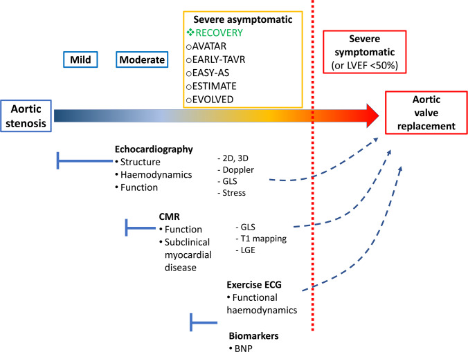
This review focuses on the available data regarding the utility of advanced left ventricular (LV) imaging in aortic stenosis (AS) and its potential impact for optimising the timing of aortic valve replacement. Ejection fraction is currently the only LV parameter recommended to guide intervention in AS. The cut-off value of 50%, recommended for decision-making in asymptomatic patients with AS, is currently under debate. Several imaging parameters have emerged as predictors of disease progression and clinical outcomes in this setting. Global longitudinal LV strain by speckle tracking echocardiography is useful for risk stratification of asymptomatic patients with severe AS and preserved LV ejection fraction. Its prognostic value was demonstrated in these patients, but further work is required to define the best thresholds to aid the decision-making process. The assessment of myocardial fibrosis is the most studied application of cardiac magnetic resonance in AS. The detection of replacement fibrosis by late gadolinium enhancement offers incremental prognostic information in these patients. Clinical implementation of this technique to optimise the timing of aortic valve intervention in asymptomatic patients is currently tested in a randomised trial. The use of T1 mapping techniques can provide an assessment of interstitial myocardial fibrosis and represents an expanding field of interest. However, convincing data in patients with AS is still lacking. All these imaging parameters have substantial potential to influence the management decision in patients with AS in the future, but data from randomised clinical trials are awaited to define their utility in daily practice.

Keywords: advanced cardiac imaging; aortic stenosis; cardiac magnetic resonance (CMR) imaging; echocardiography.


Conflict of interest statement

Competing interests: BAP has received research support and lecture honoraria from General Electric Healthcare.

Figures

Figure 1
Figure 1
Consequences of very severe calcific aortic stenosis (A) in a patient in their 60s without a history of systemic hypertension. Peak transvalvular gradient was 127 mm Hg (mean gradient of 84 mm Hg) (B) at a blood pressure of 120/70 mm Hg, with an estimated LV systolic pressure of 247 mm Hg and a calculated aortic valve area of 0.6 cm2. As a result of severe pressure overload, there was significant concentric LV hypertrophy (A) with an indexed LV mass of 130 g/m2 and a relative wall thickness of 0.55. Global systolic LV function was preserved (ejection fraction of 65%) while systolic myocardial velocities measured by tissue Doppler imaging were significantly reduced (septal s’ 5.2 cm/s)(D), indicating LV longitudinal dysfunction. There was significant LV diastolic dysfunction: impaired relaxation with a septal e’ of 4.8 cm/s (D) and increased filling pressure—pseudonormal mitral inflow (C) with elevated E/e' ratio of 16 and a moderately dilated left atrium. LV, left ventricular.
Figure 2
Figure 2
LV global longitudinal strain measured by speckle tracking echocardiography in a patient with severe aortic stenosis before (A) and 1 month after TAVI (B). Before TAVI, the LVEF was 30% and the mean transvalvular gradient was 70 mm Hg. The GLS was severely reduced: −5.7%. Systolic lengthening is present in the basal and mid-segments of the lateral wall. Angiography revealed no significant coronary artery disease. One month after TAVI, LVEF increased to 55% while GLS significantly improved: −13.3%. GLS, global longitudinal strain; LV, left ventricle; LVEF, LV ejection fraction; TAVI, transcatheter aortic valve implantation.
Figure 3
Figure 3
LV mechanical dispersion assessed by speckle-tracking echocardiography in the four-chamber view in two patients with severe aortic stenosis and preserved LV ejection fraction. White horizontal arrows indicate contraction duration, defined as the time from QRS onset on the ECG to peak negative longitudinal strain. Mechanical dispersion is defined as the SD of contraction duration from all LV segments. (A) Low value of mechanical dispersion indicating a homogeneous contraction pattern. (B) Pronounced mechanical dispersion in a patient with significant LV hypertrophy and complex ventricular arrythmia. LV: Left ventricular.
Figure 4
Figure 4
LGE on CMR imaging. Panels A and B are short axis and three-chamber views of a patient with aortic stenosis. The red arrows indicate focal, non-ischaemic (mid-wall) LGE in the basal inferolateral wall. The dotted blue arrow in panel A indicates the left ventricular outflow tract, where caution must be taken in interpreting contrast enhancement due to partial volume effects. in contrast, panels C and D are the same views of a different patient with a myocardial infarction involving the same segments (orange arrows). Note the transmural pattern and wall thinning. LGE, Late gadolinium enhancement; CMR, cardiac magnetic resonance.
Figure 5
Figure 5
Proposed schematic integrating multi-modality LV assessments in AS. The current paradigm remains one of aortic valve replacement when AS is severe and symptomatic, or if there is overt evidence of impaired LV function (reduced ejection fraction). Several randomised controlled trials are assessing early intervention in severe asymptomatic AS (orange box). Of these, only RECOVERY has been completed, demonstrating a clinical benefit with early intervention in a population with very severe AS, a large proportion of which were bicuspid. EVOLVED is the only trial utilising imaging to select a subset of asymptomatic patients for early intervention. The other trials assess if early intervention could be beneficial in this setting, regardless of adjunctive risk stratification tools. Echocardiography remains, and should remain, the first-line investigation to assess AS and its effect on the LV. However, CMR adds incremental information regarding subclinical myocardial disease (fibrosis) via tissue characterisation that cannot be detected with echocardiography; these pathological changes are often present before ejection fraction falls and have prognostic implications. Other indirect assessments of LV function in response to the fixed afterload induced by AS include exercise ECG and biomarkers such as BNP. AS, aortic stenosis; BNP, B-type natriuretic peptide; CMR, cardiac magnetic resonance; RECOVERY, Randomized Comparison of Early Surgery Versus Conventional Treatment in Very Severe Aortic Stenosis, EVOLVED, Early Valve Replacement Guided by Biomarkers of LV Decompensation in Asymptomatic Patients with Severe AS; GLS, global longitudinal strain; LV, left ventricular.

References

    1. Nkomo VT, Gardin JM, Skelton TN, et al. . Burden of valvular heart diseases: a population-based study. The Lancet 2006;368:1005–11. 10.1016/S0140-6736(06)69208-8 - DOI - PubMed
    1. Tastet L, Tribouilloy C, Maréchaux S, et al. . Staging cardiac damage in patients with asymptomatic aortic valve stenosis. J Am Coll Cardiol 2019;74:550–63. 10.1016/j.jacc.2019.04.065 - DOI - PubMed
    1. Grossman W, Jones D, McLaurin LP. Wall stress and patterns of hypertrophy in the human left ventricle. J Clin Invest 1975;56:56–64. 10.1172/JCI108079 - DOI - PMC - PubMed
    1. Chin CWL, Everett RJ, Kwiecinski J, et al. . Myocardial fibrosis and cardiac decompensation in aortic stenosis. JACC Cardiovasc Imaging 2017;10:1320–33. 10.1016/j.jcmg.2016.10.007 - DOI - PMC - PubMed
    1. Salcedo EE, Korzick DH, Currie PJ, et al. . Determinants of left ventricular hypertrophy in patients with aortic stenosis. Cleve Clin J Med 1989;56:590–6. 10.3949/ccjm.56.6.590 - DOI - PubMed

Publication types

MeSH terms