Review
. 2020 Mar 16;133(6):jcs228031.
doi: 10.1242/jcs.228031.

Scaling up single-cell mechanics to multicellular tissues - the role of the intermediate filament-desmosome network

Affiliations
Review

Scaling up single-cell mechanics to multicellular tissues - the role of the intermediate filament-desmosome network

Joshua A Broussard et al. J Cell Sci. .

Abstract

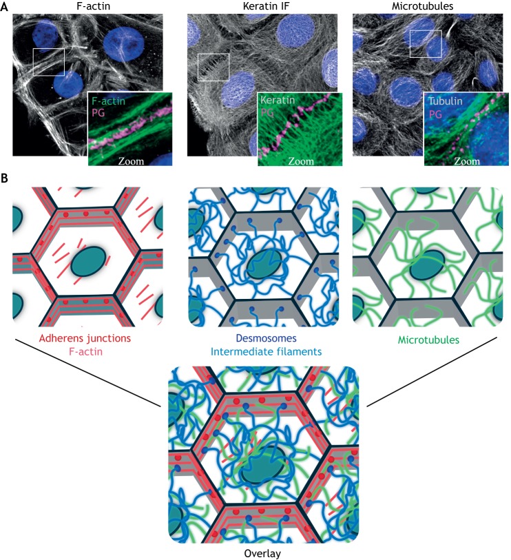
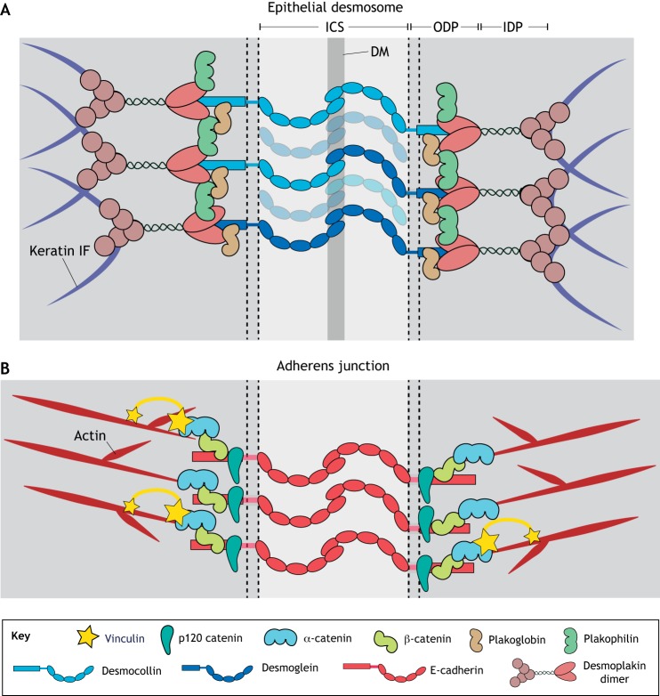
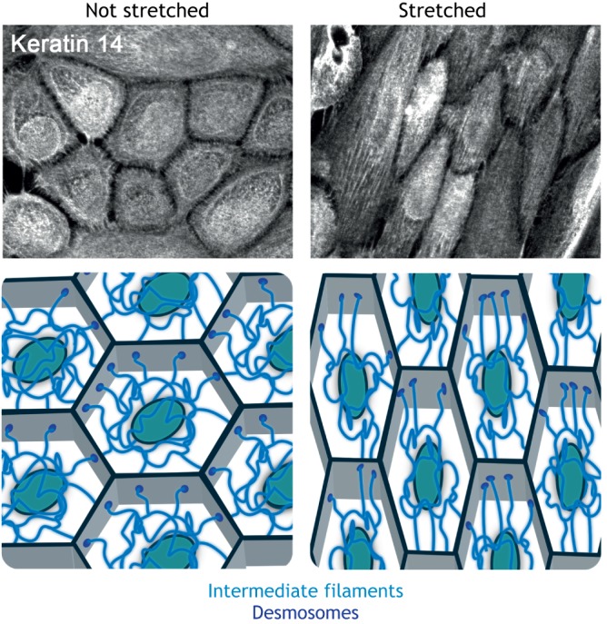
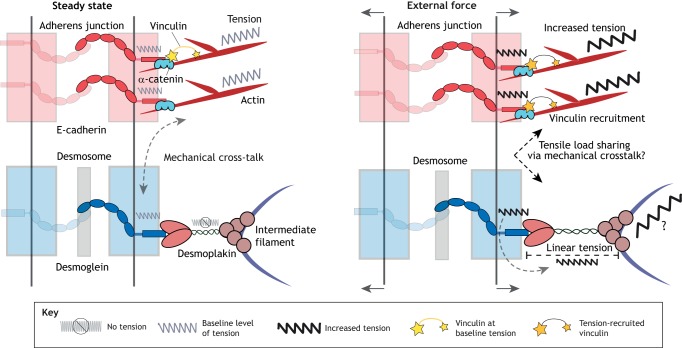
Cells and tissues sense, respond to and translate mechanical forces into biochemical signals through mechanotransduction, which governs individual cell responses that drive gene expression, metabolic pathways and cell motility, and determines how cells work together in tissues. Mechanotransduction often depends on cytoskeletal networks and their attachment sites that physically couple cells to each other and to the extracellular matrix. One way that cells associate with each other is through Ca2+-dependent adhesion molecules called cadherins, which mediate cell-cell interactions through adherens junctions, thereby anchoring and organizing the cortical actin cytoskeleton. This actin-based network confers dynamic properties to cell sheets and developing organisms. However, these contractile networks do not work alone but in concert with other cytoarchitectural elements, including a diverse network of intermediate filaments. This Review takes a close look at the intermediate filament network and its associated intercellular junctions, desmosomes. We provide evidence that this system not only ensures tissue integrity, but also cooperates with other networks to create more complex tissues with emerging properties in sensing and responding to increasingly stressful environments. We will also draw attention to how defects in intermediate filament and desmosome networks result in both chronic and acquired diseases.

Keywords: Cadherin; Cell–cell adhesion; Cytoskeleton; Desmosome; Intermediate filaments; Mechanotransduction.


Conflict of interest statement

Competing interestsThe authors declare no competing or financial interests.

Figures

Fig. 1.
Fig. 1.
Architecture of the three main cytoskeletal systems. (A) Immunofluorescence staining shows the organization of the F-actin, keratin IF and microtubule cytoskeletons in human epidermal keratinocytes. Plakoglobin (PG) is used to show regions of cell–cell contact and nuclei are shown in blue with DAPI. (B) Schematic representations of the indicated filamentous cytoskeletons and associated cell–cell adhesive complexes, as well as an overlay to illustrate their interconnected nature. Gray outlines represent cell–cell junctional areas and ovals represent nuclei. Images supplied by the laboratory of K.J.G.
Fig. 2.
Fig. 2.
Major desmosome and adherens junction components. (A) Transmembrane desmosomal cadherins form extracellular interactions between adjacent cells. The cytoplasmic domains of these cadherins bind to the armadillo proteins plakophilin and plakoglobin and together bind desmoplakin, which anchors the desmosome to the IF cytoskeleton. (B) Adherens junctions contain classical transmembrane cadherins that facilitate cell–cell interactions owing to interactions between their extracellular domains. Intracellularly, classical cadherins interact with the armadillo protein β-catenin and catenin family proteins, including p120 catenin and α-catenin. Linkage to the actin cytoskeleton is mediated through α-catenin, as well as vinculin, which is recruited to the junction under tension. ICS, intercellular space; IDP, inner dense plaque; ODP, outer dense plaque; DM, dense midline.
Fig. 3.
Fig. 3.
Changes to the IF cytoskeleton under external force. Mechanical stretch induces alignment of the IF network. Neonatal epidermal keratinocytes were incubated with 1.2 mM Ca2+-containing medium overnight to induce the formation of robust cell–cell junctions. Cell monolayers were then subjected to cyclical stretch for 24 h, fixed, and stained for keratin 14 to demonstrate reorganization of the IF cytoskeleton. Control cells were not stretched. Images supplied by the laboratory of K.J.G. A schematic representation of the reorganization of the IF cytoskeleton that occurs upon application of mechanical stimulation is also shown.
Fig. 4.
Fig. 4.
Contributions of junctional and cytoskeletal proteins to cellular mechanics. At steady state (left), F-actin and the associated classical cadherins are under a baseline level of tension at the adherens junction. In addition, at steady state, desmosomal cadherins are under low tension, while desmoplakin experiences little to no tension. It is not known why the low tensile forces on desmosomal cadherins fail to be propagated to the IF cytoskeleton, but this could be explained through a mechanical crosstalk with the tensile F-actin-based adhesive system. When an external force is applied (right), the tensile load is shared between adherens junctions and desmosomes. Increased tension is ‘felt’ by the classical cadherins and F-actin at the adherens junction, leading to increased recruitment of vinculin, thereby facilitating mechanotransduction. When an external force is applied, the desmosomal cadherins propagate tensile forces to desmoplakin and potentially the IF network in order for cells and tissues to resist large mechanical loads.

References

    1. Ai-Jassar C., Bikker H., Overduin M. and Chidgey M (2013). Mechanistic basis of desmosome-targeted diseases. J. Mol. Biol. 425, 4006-4022. 10.1016/j.jmb.2013.07.035 - DOI - PMC - PubMed
    1. Alami N. H., Jung P. and Brown A (2009). Myosin Va increases the efficiency of neurofilament transport by decreasing the duration of long-term pauses. J. Neurosci. 29, 6625-6634. 10.1523/JNEUROSCI.3829-08.2009 - DOI - PMC - PubMed
    1. Ananthakrishnan R. and Ehrlicher A (2007). The forces behind cell movement. Int. J. Biol. Sci. 3, 303-317. 10.7150/ijbs.3.303 - DOI - PMC - PubMed
    1. Asimaki A., Syrris P., Wichter T., Matthias P., Saffitz J. E. and Mckenna W. J. (2007). A novel dominant mutation in plakoglobin causes arrhythmogenic right ventricular cardiomyopathy. Am. J. Hum. Genet. 81, 964-973. 10.1086/521633 - DOI - PMC - PubMed
    1. Baddam S. R., Arsenovic P. T., Narayanan V., Duggan N. R., Mayer C. R., Newman S. T., Abutaleb D. A., Mohan A., Kowalczyk A. P. and Conway D. E. (2018). The desmosomal cadherin desmoglein-2 experiences mechanical tension as demonstrated by a FRET-based tension biosensor expressed in living cells. Cells 7, E66 10.3390/cells7070066 - DOI - PMC - PubMed

Publication types