

Microbiological and scanning electron microscopic evaluation of epidural catheters
- PMID: 32179623
- PMCID: PMC8408583
- DOI: 10.1136/rapm-2019-101180
Microbiological and scanning electron microscopic evaluation of epidural catheters
Abstract
Background: Epidural catheters are frequently colonized by gram-positive bacteria. Although the incidence of associated epidural infections is low, their consequences can be devastating. We investigated bacterial growth on epidural catheters by quantitative bacterial culture and scanning electron microscopy (SEM) in order to explore the patterns of epidural catheter colonization.
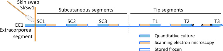
Methods: 28 patients undergoing major abdominal surgery with thoracic epidurals (treatment ≥72 hours) were studied. Before the removal of the catheter, the skin surrounding the insertion site was swabbed. The entire catheter was divided into extracorporeal, subcutaneous, and tip segments. Skin swabs and catheter segments were quantitatively cultured, bacterial species were identified, and SEM was performed on four selected catheters.
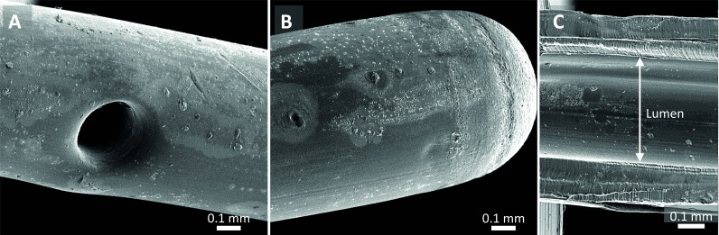
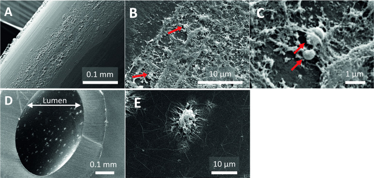
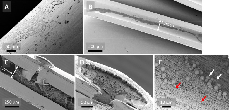
Results: 27 of 28 catheters were included. The percentages of positive cultures were: skin swab 29.6%, extracorporeal segments 11.1%, subcutaneous segments 14.8%, and tip segments 33.3%. One patient was diagnosed with a catheter-associated infection. Staphylococcus epidermidis was cultured from the skin and the catheter extracorporeal, subcutaneous, and tip segments. SEM of this catheter showed bacteria-like and intraluminal host cell-like structures. SEM of two other catheters showed intraluminal fibrin networks in their tip segments.
Conclusions: We present the first SEM pictures of an epidural catheter with a bacterial infection. Bacterial growth developed from the skin to the tip of this catheter, indicating the skin as a primary source of infection. By SEM, catheters with low levels of bacterial growth demonstrated an intraluminal fibrous network which possibly plays a role in catheter obstruction.
Keywords: neuraxial blocks: continuous techniques; neuraxial blocks: epidural; regional anesthesia.
© American Society of Regional Anesthesia & Pain Medicine 2020. Re-use permitted under CC BY. Published by BMJ.
Conflict of interest statement
Competing interests: None declared.
Figures
Similar articles
-
Bacterial colonization of epidural catheters used for short-term postoperative analgesia: microbiological examination and risk factor analysis.Anesthesiology. 2008 Jan;108(1):130-7. doi: 10.1097/01.anes.0000296066.79547.f3. Anesthesiology. 2008. PMID: 18156891
-
Culture of bacteria from lumbar and caudal epidural catheters used for postoperative analgesia in children.Reg Anesth. 1997 Sep-Oct;22(5):428-31. doi: 10.1016/s1098-7339(97)80028-4. Reg Anesth. 1997. PMID: 9338903 Clinical Trial.
-
Frequency of colonization and isolated bacteria from the tip of epidural catheter implanted for postoperative analgesia.Braz J Anesthesiol. 2015 May-Jun;65(3):200-6. doi: 10.1016/j.bjane.2014.05.015. Epub 2015 Mar 30. Braz J Anesthesiol. 2015. PMID: 25925032
-
[Severe infections after epidural catheterization].Ugeskr Laeger. 1998 May 25;160(22):3202-6. Ugeskr Laeger. 1998. PMID: 9621797 Review. Danish.
-
Why do thoracic epidurals fail? A literature review on thoracic epidural failure and catheter confirmation.World J Crit Care Med. 2024 Sep 9;13(3):94157. doi: 10.5492/wjccm.v13.i3.94157. eCollection 2024 Sep 9. World J Crit Care Med. 2024. PMID: 39253309 Free PMC article. Review.
Cited by
-
Scanning Electron Microscope Examination as an Alternative to Classical Microbiology in the Diagnostics of Catheter-Related Sepsis?Int J Environ Res Public Health. 2023 Mar 13;20(6):5028. doi: 10.3390/ijerph20065028. Int J Environ Res Public Health. 2023. PMID: 36981937 Free PMC article.
-
Epidural infections, bacteriostatic drug effects and technical strategies for prevention.Reg Anesth Pain Med. 2022 Feb;47(2):128-130. doi: 10.1136/rapm-2021-102517. Epub 2021 Jun 11. Reg Anesth Pain Med. 2022. PMID: 34117104 Free PMC article. No abstract available.
-
Clinical Application of Different Doses of Hydromorphone Slow-Release Analgesia in Lumbar Fusion in Elderly Patients.Pain Ther. 2024 Oct;13(5):1219-1233. doi: 10.1007/s40122-024-00632-3. Epub 2024 Jul 12. Pain Ther. 2024. PMID: 38995609 Free PMC article.
References
MeSH terms
LinkOut - more resources
Full Text Sources