. 2020 Mar;8(1):e000317.
doi: 10.1136/jitc-2019-000317.

Compound kushen injection relieves tumor-associated macrophage-mediated immunosuppression through TNFR1 and sensitizes hepatocellular carcinoma to sorafenib

Affiliations

Compound kushen injection relieves tumor-associated macrophage-mediated immunosuppression through TNFR1 and sensitizes hepatocellular carcinoma to sorafenib

Yang Yang et al. J Immunother Cancer. 2020 Mar.

Erratum in

Abstract

Background: There is an urgent need for effective treatments for hepatocellular carcinoma (HCC). Immunotherapy is promising especially when combined with traditional therapies. This study aimed to investigate the immunomodulatory function of an approved Chinese medicine formula, compound kushen injection (CKI), and its anti-HCC efficiency in combination with low-dose sorafenib.

Methods: Growth of two murine HCC cells was evaluated in an orthotopic model, a subcutaneous model, two postsurgical recurrence model, and a tumor rechallenge model with CKI and low-dose sorafenib combination treatment. In vivo macrophage or CD8+ T cell depletion and in vitro primary cell coculture models were used to determine the regulation of CKI on macrophages and CD8+ T cells.

Results: CKI significantly enhanced the anticancer activity of sorafenib at a subclinical dose with no obvious side effects. CKI and sorafenib combination treatment prevented the postsurgical recurrence and rechallenged tumor growth. Further, we showed that CKI activated proinflammatory responses and relieved immunosuppression of tumor-associated macrophages in the HCC microenvironment by triggering tumor necrosis factor receptor superfamily member 1 (TNFR1)-mediated NF-κB and p38 MAPK signaling cascades. CKI-primed macrophages significantly promoted the proliferation and the cytotoxic ability of CD8+ T cells and decreased the exhaustion, which subsequently resulted in apoptosis of HCC cells.

Conclusions: CKI acts on macrophages and CD8+ T cells to reshape the immune microenvironment of HCC, which improves the therapeutic outcomes of low-dose sorafenib and avoids adverse chemotherapy effects. Our study shows that traditional Chinese medicines with immunomodulatory properties can potentiate chemotherapeutic drugs and provide a promising approach for HCC treatment.

Keywords: gastroenterology; immunology; pharmacology; tumours.


Conflict of interest statement

Competing interests: None declared.

Figures
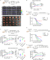
Figure 1
Figure 1
Compound kushen injection (CKI) combined with low-dose sorafenib suppressed liver cancer growth, reduced postsurgical recurrence and the growth of rechallenged tumors in mice. (A) The schedule of orthotopic liver cancer model treatment and imaging (above panel). Representative bioluminescence images of mice with orthotopic Hepa1-6 tumor burden (middle panel). Mice were treated daily with CKI intraperitoneally (150 µL), sorafenib intragastrically (10 mg/kg), CKI combined with sorafenib, or the vehicle (n=6 per group). Representative photographs of liver tumors after the indicated treatments; the red dotted lines indicate the tumor regions (below panel). (B) Relative luminescence changes of liver tumors depicted in (A) during the treatment. (C) The schedule of subcutaneous liver cancer model treatment and tumor measurement (above panel). A total of 2×10⁶ LPC-H12 or Hepa1-6 cells were separately injected into the left flanks of C57BL/6 mice (n=6–8 per group). Mice were treated daily with the indicated drugs in accordance with the orthotopic model. Tumor growth and tumor weight were measured. (D) The schedule of postsurgical cancer treatment and recurrence observation (above panel). Percentage of recurrence-free survival of mice was shown (vehicle, n=11; CKI, n=12; sorafenib, n=12; CKI+sorafenib, n=10). (E) The schedule of postsurgical cancer treatment and recurrence observation (above panel). Percentage of recurrence-free survival of mice was shown (vehicle, n=11; CKI, n=12; sorafenib, n=11; CKI+sorafenib, n=14). (F) The schedule of postsurgical tumor rechallenge model treatment and measurement (above panel). The volume of rechallenged tumor in mice was measured (n=7). Data are presented as means±SEM. NS, p>0.05; *p<0.05; **p<0.01; ***p<0.001.
Figure 2
Figure 2
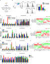
Compound kushen injection (CKI) combined with low-dose sorafenib decreased the distribution of M2-TAMs, increased the ratio of M1-TAMs and CD8+ T cells in the tumor microenvironment. (A) The proportion of tumor-infiltrating immune cells in mice tumor tissues after treatment was quantified by FACS. (B) Representative flow cytometry gating images show the percentages of CD8+ T cells, CD4+ T cells, M1-TAMs and M2-TAMs in mice orthotopic Hepa1-6 tumor tissues. (C) The expression of function exhaustion markers (Lag-3, PD-1, TIGIT and Tim-3) on CD8+ T cells after treatment was detected by flow cytometry. (D) Representative immunohistochemistry of INOS, Arg-1, CD8, cleaved caspase-3 and TUNEL stain in LPC-H12 tumor sections was shown. Data are presented as means±SEM. *p<0.05; **p<0.01; ***p<0.001. Arg-1, arginase 1; INOS, Induced nitric oxide synthase; Lag 3, lymphocyte activation gene 3; PD-1, programmed cell death protein 1; TAMs, tumor-associated macrophages; TIGIT, T-cell immunoreceptor with Ig and ITIM domains; Tim-3: T-cell immunoglobulin and mucin-domain containing-3; TUNEL, terminaldeoxynucleotidyl transferase mediated dUTP nck-end labeling.
Figure 3
Figure 3
Depletion of macrophages or CD8+ T cells abolished the anti-hepatocellular carcinoma (HCC) effect of compound kushen injection (CKI) combined with low-dose sorafenib. (A) C57BL/6 mice (n=7–8) bearing LPC-H12 subcutaneous tumor were injected with neutralizing anti-CD8 antibody to deplete CD8+ T cells. After drug treatments, tumor growth, tumor weight, and body weight were measured. (B) The proportion of CD4+ T, CD8+ T, M1-TAMs, and M2-TAMs in CD8+ T cell-depleted tumors were determined by FACS. (C) C57BL/6 mice (n=7) were planted with LPC-H12 tumor cells subcutaneously and then treated with clodronate liposomes to deplete macrophages. After the indicated treatments, tumor growth, tumor weight, and body weight were measured. (D) The proportion of CD4+ T, CD8+ T, M1-TAMs, and M2-TAMs in macrophage-depleted tumors were determined by FACS. (E) Schematic diagram of macrophage replenishment in C57BL/6 with macrophage depletion by clodronate liposome. (F) The tumor growth and tumor weight were measured in different groups with the indicated treatments after macrophage replenishment. (G) The proportion of CD4+ T and CD8+ T cells in macrophage replenishment tumor tissues after treatments. (H) Immunohistochemical staining of INOS, Arg-1, and CD8 in CD8+ T cell-depleted LPC-H12 subcutaneous tumors. (I) Immunohistochemical staining of INOS, Arg-1, and CD8 in macrophage-depleted LPC-H12 subcutaneous tumors. Data are presented as means±SEM. NS, p>0.05; *p<0.05; **p<0.01; ***p<0.001. Arg-1, arginase 1; INOS, induced nitric oxide synthase.
Figure 4
Figure 4
Compound kushen injection (CKI) promoted tumor-educated macrophage polarization to M1 status, enhanced proinflammatory function and reduced anti-inflammatory function. (A) Schematic diagram of bone marrow-derived macrophages (BMDMs) (M0) different polarization status after treatment conditioned medium (CM) for 24 hours (Mhepa1-6), 100 ng/mL LPS and 50 ng/mL IFN-γ for 12 hours (M1) or 10 ng/mL IL-4 and 10 ng/mL IL-13 for 24 hours (M2) followed by CKI treatment. (B) Mhepa1-6 were incubated with different concentrations of CKI for 12 hours. The proportion of M1 and M2 macrophages was determined by FACS. (C) The expressions of M1 (proinflammation) and M2 (anti-inflammation) macrophage markers in Mhepa1-6 with different doses of CKI treatment for 12 hours were determined by quantitative real-time PCR (qRT-PCR). (D) The contents of M1 and M2-related cytokines or proteins were measured by ELISA in CKI-primed Mhepa1-6 cell culture supernatant or cell lysates. (E and F) BMDMs (M0) were polarized to M1 status and treated with different doses of CKI for 12 hours. The expression of proinflammation and anti-inflammation genes was determined by qRT-PCR (E) or ELISA (F). (G and H) BMDMs (M0) were polarized to M2 status and treated with different doses of CKI for 24 hours. The expression of proinflammation and anti-inflammation genes was determined by qRT-PCR (G) or ELISA (H). (I) Tumor-associated macrophages were sorted from LPC-H12 subcutaneous tumors in mice after the indicated treatments. The mRNA levels of proinflammation and anti-inflammation genes were detected by qRT-PCR. Data are presented as means±SEM. *p<0.05; **p<0.01; ***p<0.001.
Figure 5
Figure 5
Compound kushen injection (CKI)-primed macrophages improved the proliferation and tumor-killing ability of CD8+ T cells, and relieved exhaustion. (A) Schematic diagram of CKI-primed macrophage and CD8+ T cell coculture system (macrophages: CD8+ T ratio=5:1, CD8+ T number=2×10⁵) with anti-CD3 (2.5 µg/mL) and anti-CD28 (5 µg/mL) stimulation for 72 hours. (B) CD8+ T cells were labeled with carboxyfluorescein succinimidyl amino ester (CFSE) and cocultured with CKI-primed macrophages (Mhepa1-6, M1 and M2) for 72 hours. Representative plots of CFSE signal intensity of CD8+ T cells were described for CD8+ T stimulated by CD3/CD28 antibody and cocultured with vehicle or CKI-primed macrophages (left panel). The quantification of proliferated CD8+ T cells was calculated in different groups (right panel). (C) The expression of cytotoxic and exhausted markers in CD8+ T cells cocultured with CKI-primed macrophages (Mhepa1-6, M1 and M2) was determined by quantitative real-time PCR (qRT-PCR). (D) The contents of cytotoxic cytokines were measured in coculture supernatants by ELISA. (E) CD8+ T cells (cocultured with CKI-primed Mhepa1-6, M1, and M2 before) were coincubated with luciferase-labeled-Hepa1-6 cells for 24 hours (CD8+T: Hepa1-6 ratio=12:1, total cells number=5000). The lysis degree of Hepa1-6 tumor cells was monitored by fluorescence values. (F) CD8+ T cells were sorted from LPC-H12 subcutaneous tumors in mice with the indicated treatments. The mRNA levels of cytotoxic and exhausted genes in CD8+ T cells were determined by qRT-PCR. Data are presented as means±SEM. *p<0.05; **p<0.01; ***p<0.001.
Figure 6
Figure 6
Compound kushen injection (CKI) facilitated TNFR1 complex interaction and NF-κB p65 and MAPK p38 signaling cascade to play a proinflammatory function. (A) Tumor-associated macrophages were FACS sorted from LPC-H12 subcutaneous tumors in mice with the indicated treatments, and the mRNA levels of proinflammatory polarization-related receptors genes were detected by quantitative real-time PCR (qRT-PCR). (B) The expression of proinflammatory polarization-related receptors in Mhepa1-6 with 0.66 mg/mL CKI incubation for 12 hours was measured. (C) The expression of proinflammation and anti-inflammation genes in Mhepa1-6 exposed to 0.66 mg/mL CKI for 12 hours with TNFR1 blockade (R7050, 10 µM) was measured. (D) Cell surface level of TNFR1 in 0.66 mg/mL CKI-primed Mhepa1-6 was measured by FACS. (E) Mhepa1-6 cells were treated with 0.66 mg/mL CKI for 12 hours, and the binding between TNFR1 with TRADD, RIP1 and TRAF2 was determined by coimmunoprecipitation. (F) Mhepa1-6 cell were exposed to 0.66 mg/mL CKI for 12 hours and 10 ng/mL TNF-α for different time intervals, and the protein expression was determined by western blotting. (G) Mhepa1-6 were treated with 25 µg/mL anti-TNFR1 antibody (left panel) or 10 µM R7050 (right panel) for 4 hours before 0.66 mg/mL CKI treatment for another 12 hours, then stimulated by 10 ng/mL TNF-α for 30 min. The amount of TRAF2, phosphorylated and total TAK1, P65 and P38 expression were quantified by western blotting. (H) Mice with subcutaneous LPC-H12 tumor burden were treated with CKI and sorafenib daily (CKI, 150 µL; sorafenib, 10 mg/kg), anti-TNFR1 antibody (100 µg per mouse, every 4 days), R-7050 (10 mg/mL, every 3 days) or the vehicle (n=7 per group). Tumor growth and tumor weight were measured. Data are presented as means±SEM. NS, p>0.05; *p<0.05; **p<0.01; ***p<0.001. TNF-α, tumor necrosis factor α; TNFR1, TNF receptor superfamily member 1.
Figure 7
Figure 7
Schematic depiction of the reconstruction of the hepatocellular carcinoma (HCC) microenvironment by compound kushen injection (CKI) and its therapeutic effects when combined with low-dose sorafenib. INOS, induced nitric oxidesynthase; IFN-β, interferon-β; IL, interleukin; Lag-3, lymphocyte-activation gene 3; Tim-3, T-cell immunoglobulin and mucin-domain containing-3; TIGIT, T-cell immunoreceptor with Ig and ITIM domains; TNF-α, tumor necrosis factor α.

Similar articles

Cited by

References

    1. Dutta R, Mahato RI. Recent advances in hepatocellular carcinoma therapy. Pharmacol Ther 2017;173:106–17. 10.1016/j.pharmthera.2017.02.010 - DOI - PMC - PubMed
    1. Villanueva A. Hepatocellular carcinoma. N Engl J Med 2019;380:1450–62. 10.1056/NEJMra1713263 - DOI - PubMed
    1. Bray F, Ferlay J, Soerjomataram I, et al. . Global cancer statistics 2018: GLOBOCAN estimates of incidence and mortality worldwide for 36 cancers in 185 countries. CA Cancer J Clin 2018;68:394–424. 10.3322/caac.21492 - DOI - PubMed
    1. Llovet JM, Zucman-Rossi J, Pikarsky E, et al. . Hepatocellular carcinoma. Nat Rev Dis Primers 2016;2:16018 10.1038/nrdp.2016.18 - DOI - PubMed
    1. Llovet JM, Hernandez-Gea V. Hepatocellular carcinoma: reasons for phase III failure and novel perspectives on trial design. Clin Cancer Res 2014;20:2072–9. 10.1158/1078-0432.CCR-13-0547 - DOI - PubMed

Publication types

MeSH terms