. 2020 Mar 31;117(13):7021-7029.
doi: 10.1073/pnas.1914911117. Epub 2020 Mar 16.

Oxaliplatin Pt(IV) prodrugs conjugated to gadolinium-texaphyrin as potential antitumor agents

Affiliations

Oxaliplatin Pt(IV) prodrugs conjugated to gadolinium-texaphyrin as potential antitumor agents

Grégory Thiabaud et al. Proc Natl Acad Sci U S A. .

Abstract

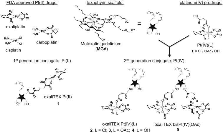
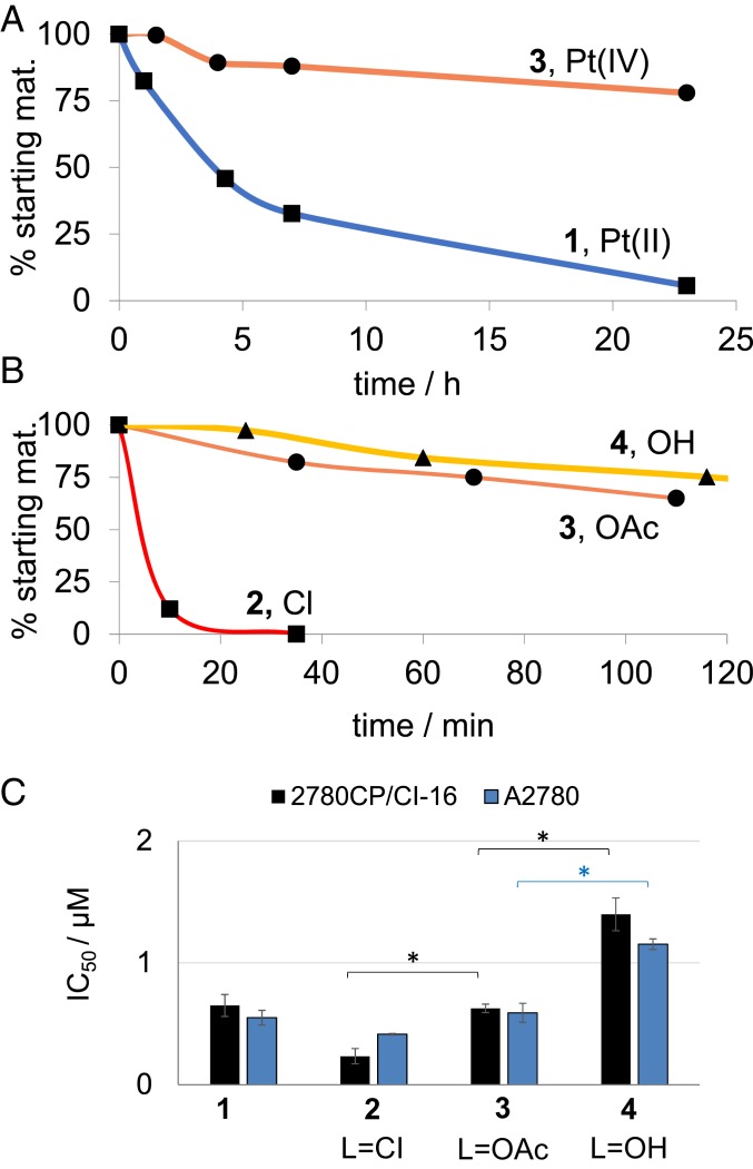
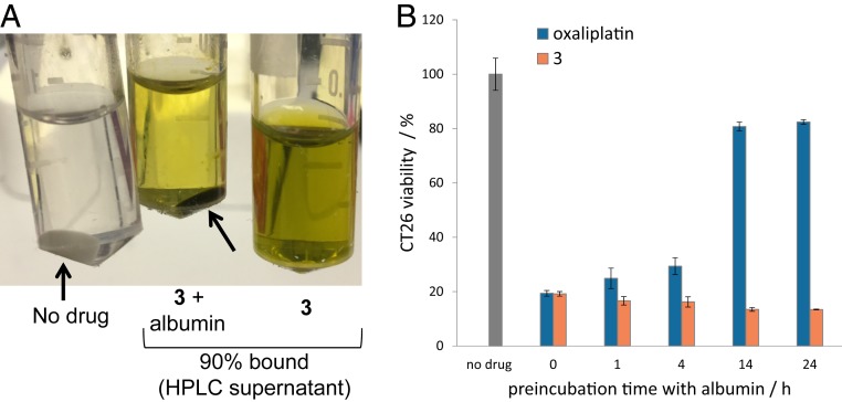
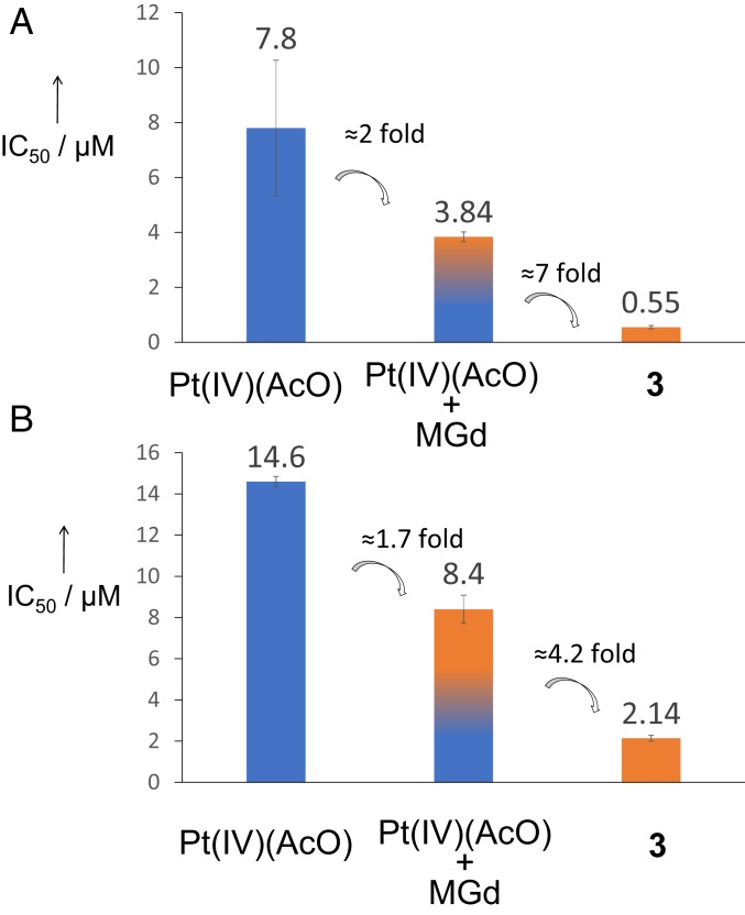
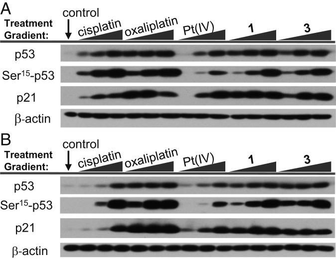
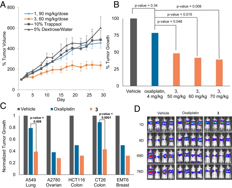
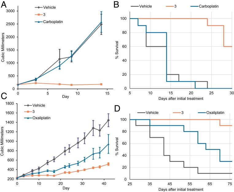
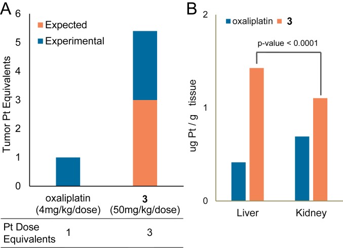
Described here is the development of gadolinium(III) texaphyrin-platinum(IV) conjugates capable of overcoming platinum resistance by 1) localizing to solid tumors, 2) promoting enhanced cancer cell uptake, and 3) reactivating p53 in platinum-resistant models. Side by side comparative studies of these Pt(IV) conjugates to clinically approved platinum(II) agents and previously reported platinum(II)-texaphyrin conjugates demonstrate that the present Pt(IV) conjugates are more stable against hydrolysis and nucleophilic attack. Moreover, they display high potent antiproliferative activity in vitro against human and mouse cell cancer lines. Relative to the current platinum clinical standard of care (SOC), a lead Gd(III) texaphyrin-Pt(IV) prodrug conjugate emerging from this development effort was found to be more efficacious in subcutaneous (s.c.) mouse models involving both cell-derived xenografts and platinum-resistant patient-derived xenografts. Comparative pathology studies in mice treated with equimolar doses of the lead Gd texaphyrin-Pt(IV) conjugate or the US Food and Drug Administration (FDA)-approved agent oxaliplatin revealed that the conjugate was better tolerated. Specifically, the lead could be dosed at more than three times (i.e., 70 mg/kg per dose) the tolerable dose of oxaliplatin (i.e., 4 to 6 mg/kg per dose depending on the animal model) with little to no observable adverse effects. A combination of tumor localization, redox cycling, and reversible protein binding is invoked to explain the relatively increased tolerability and enhanced anticancer activity seen in vivo. On the basis of the present studies, we conclude that metallotexaphyrin-Pt conjugates may have substantial clinical potential as antitumor agents.

Keywords: cancer; drug development; drug resistance; platinum prodrug; texaphyrins.


Conflict of interest statement

Competing interest statement: Since the time of the original submission, the texaphyrin conjugates described in this manuscript were licensed by The University of Texas to the IQ Global Group and planned for further development by a new for-profit company, OncoTEX Inc. J.F.A. is now employed by OncoTEX Inc., and J.L.S. now serves as a nonexecutive board member for OncoTEX Inc. The other authors declare no conflict of interest.

Figures

Fig. 1.
Fig. 1.
Structures of FDA-approved platinum drugs, the first generation oxaliTEX-Pt(II) conjugate 1, and second generation conjugates 2 to 5. The latter species were synthesized by conjugation of MGd to one or two Pt(IV) prodrugs.
Fig. 2.
Fig. 2.
(A) Hydrolysis kinetic profiles determined in PBS (pH 7) at 37 °C for 1 vs. 3. (B) Reduction kinetic profiles of conjugates 2 to 4 with L = OH (triangles), L = OAc (circles), and L = Cl (squares) at 20 μM in the presence of GSH (66 μM) in PBS (pH 7) at 37 °C. Reverse phase (RP)-HPLC monitoring: detector set at 470 nm. (C) Antiproliferative activities of 1 to 4 against A2780 (blue) and 2780CP/Cl-16 (black) seen following a 5-d incubation time with the indicated platinum species. Error bars represent SD. *P values from t test (two tailed, unpaired) < 0.05.
Fig. 3.
Fig. 3.
(A) Albumin pellets (arrows) obtained in the absence (Left) or presence of 3 (0.6 mM; Center); also shown is a solution of 3 (0.6 mM) in just water (Right). The % bound of 3 to albumin was determined by HPLC analysis. (B) Antiproliferative activities against CT26 cells of oxaliplatin (1.7 μM) and 3 (4 μM) seen on preincubation with albumin (40 mg/mL). Note that cells were exposed to the Pt agent in question for 5 d.
Fig. 4.
Fig. 4.
Antiproliferative activities of Pt(IV)(AcO), of Pt(IV)(AcO) in combination with MGd (1 equivalent), and of conjugate 3 against (A) ovarian A2780 and (B) lung A549 cells; 5-d incubation time. Note that IC50(MGd) >> IC50(Pt[IV][AcO]) in both cell lines. Error bars represent SD.
Fig. 5.
Fig. 5.
p53 pathway activation of (A) A2780 cisplatin-sensitive and (B) 2780CP/Cl-16 cisplatin-resistant ovarian cancer cells treated with variable concentrations of platinum agent.
Fig. 6.
Fig. 6.
In vivo efficacy of 3, 1, and oxaliplatin in mice bearing s.c. A549 xenograft tumors (A and B). Error bars represent SD, and P value between 1 and 3 = 0.006. (C) In vivo efficacy of 3 (70 mg/kg per dose on days 1, 5, 9, 13) vs. oxaliplatin (4 mg/kg per dose on days 1, 5, 9, 13) in various s.c. xenograft tumor (A549, A2780, HCT116) and syngeneic tumor (CT26, EMT6) models. The study end point for the A549 model was 30 d. The study end point for all other models was the day at which the vehicle-treated mice reached maximum tumor burden. (D) Bioluminescent monitoring of orthotopic xenografts of A549 lung cancer cells expressing the luciferase gene. Mice were subject to i.v. administrations of 3 (at 70 mg/kg per dose) and oxaliplatin (4 mg/kg per dose) on days 9, 13,17, and 21. Bioluminescent images were acquired 10 min after intraperitoneal injection with luciferin. For AC, data represent an average for each study arm. SI Appendix has details. This includes a graph showing quantification of the luminescent signal over time corresponding to D (SI Appendix, Fig. S35).
Fig. 7.
Fig. 7.
(A) Tumor growth delay and (B) Kaplan–Meier curves of mice bearing 0253 ovarian PDX tumors. (C) Tumor growth delay and (D) Kaplan–Meier curves of mice bearing 0069 colon PDX tumors. In both studies, mice were administered 70 mg/kg per dose of 3 i.v. on days 0, 4, 8, and 12 as scheduled. For the 0253 ovarian, mice i.v. received 30 mg/kg per dose of carboplatin on days 0, 4, 8, 12, 16, and 20. For 0069 colon mice, 6 mg/kg per dose of oxaliplatin was administered i.v. on days 0, 4, and 8. For A and C, the data represent an average for each study arm. Error bars represent SD. P value between 3 and carboplatin on day 14 was 0.0002, and P value between 3 and oxaliplatin on day 31 was 0.0001. SI Appendix has details.
Fig. 8.
Fig. 8.
(A) Intratumoral platinum content (in mice) quantified by FAAS 24 h after injection of oxaliplatin (4 mg/kg) or 3 (50 mg/kg). (B) Platinum levels in liver and kidney determined by FAAS 24 h after equimolar single i.v. injection of oxaliplatin or 3 into nontumor-bearing mice. Data represent an average for each study arm. SI Appendix has details.

References

    1. Rosenberg B., VanCamp L., Trosko J. E., Mansour V. H., Platinum compounds: A new class of potent antitumour agents. Nature 222, 385–386 (1969). - PubMed
    1. Galanski M., Jakupec M. A., Keppler B. K., Update of the preclinical situation of anticancer platinum complexes: Novel design strategies and innovative analytical approaches. Curr. Med. Chem. 12, 2075–2094 (2005). - PubMed
    1. Rabik C. A., Dolan M. E., Molecular mechanisms of resistance and toxicity associated with platinating agents. Cancer Treat. Rev. 33, 9–23 (2007). - PMC - PubMed
    1. Ozols R. F., Challenges for chemotherapy in ovarian cancer. Ann. Oncol. 17 (suppl. 5), v181–v187 (2006). - PubMed
    1. Siddik Z. H., Cisplatin: Mode of cytotoxic action and molecular basis of resistance. Oncogene 22, 7265–7279 (2003). - PubMed

Publication types