. 2020 Mar 16;10(1):4767.
doi: 10.1038/s41598-020-61759-9.

Repeatability and Reproducibility of Pancreas Volume Measurements Using MRI

Affiliations

Repeatability and Reproducibility of Pancreas Volume Measurements Using MRI

Jonathan M Williams et al. Sci Rep. .

Abstract

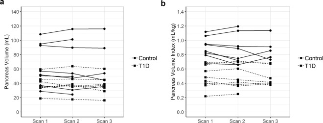
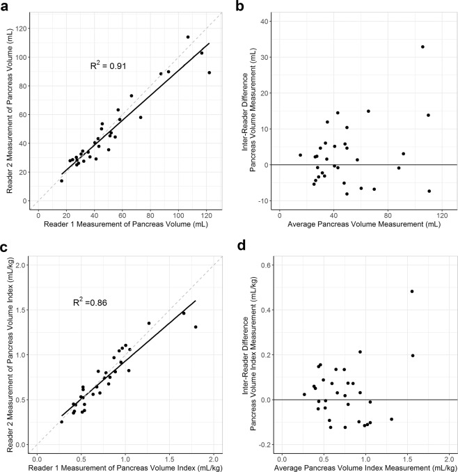
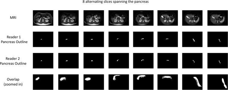
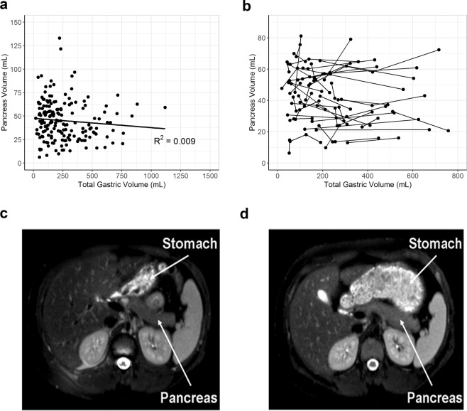
Reduced pancreas volume, as measured by non-contrast magnetic resonance imaging (MRI), is observed in individuals with newly-diagnosed type 1 diabetes (T1D) and declines over the first year after diagnosis. In this study, we determined the repeatability and inter-reader reproducibility of pancreas volume measurements by MRI. Test-retest scans in individuals with or without T1D (n = 16) had an intraclass correlation coefficient (ICC) of 0.985 (95% CI 0.961 to 0.995) for pancreas volume. Independent pancreas outlines by two board-certified radiologists (n = 30) yielded an ICC of 0.945 (95% CI 0.889 to 0.973). The mean Dice coefficient, a measurement of the degree of overlap between pancreas regions of interest between the two readers, was 0.77. Prandial state did not influence pancreatic measurements, as stomach volume did not correlate with pancreas volume. These data demonstrate that MRI measurements of pancreas volume between two readers are repeatable and reproducible with ICCs that correspond to excellent clinical significance (ICC > 0.9), are not related to changes in stomach volume, and could be a useful tool for clinical investigation of diabetes and other pancreas pathologies.


Conflict of interest statement

The authors declare no competing interests.

Figures

Figure 1
Figure 1
Measurements of pancreas volume are repeatable. Two or three repeat MRIs were performed on the same day in individuals with and without T1D (dashed and solid lines, respectively). Repeat measurements of pancreas volume (a) and pancreas volume index (b) are shown for each subject. Pancreas volume index was calculated by dividing the pancreas volume by the subject’s weight. Intraclass correlation coefficients (ICC) were reported with a 95% confidence interval.
Figure 2
Figure 2
Measurements of pancreas volume between two readers are reproducible. Two board-certified radiologists independently outlined the pancreas of 30 different individuals (31 total scans). Correlation between measurements of pancreas volume (a) and pancreas volume index (c) between the two readers is shown. Bland–Altman plots displaying the differences in measurements between readers plotted against the average measurement taken for each subject are shown for both pancreas volume (b) and pancreas volume index (d). Pancreas volume index was calculated by dividing the pancreas volume by the subject’s weight. ICCs were reported with a 95% confidence interval.
Figure 3
Figure 3
Outlines of pancreas by two readers of the same images overlap. The pancreas from a representative subject (Dice coefficient = 0.77) was outlined by two board-certified radiologists across eight alternating MRI slices of the pancreas. The overlap between the two readers’ pancreas outlines is shown in the bottom row and zoomed in for clarity. Regions of agreement are shown in bright white, while gray regions denote regions of non-overlap. The average Dice coefficient was 0.77 for the 31 scans analyzed.
Figure 4
Figure 4
Stomach volume did not affect measurements of pancreas volume. (a) Correlation between measurements of pancreas volume and total stomach volume in individuals whose MRI data sets contained the entire stomach volume within the field of view (R2 = 0.009). (b) Individuals who had longitudinal measurements of pancreas and stomach volume. Representation of an individual with low stomach volume (c) and in an apparent fed state (d).

References

    1. Chiarelli F, Verrotti A, Altobelli E, Blasetti A, Morgese G. Size of the pancreas in type I diabetic children and adolescents. Diabetes Care. 1995;18:1505–1506. doi: 10.2337/diacare.18.11.1504. - DOI - PubMed
    1. Fonseca V, Berger LA, Beckett AG, Dandona P. Size of pancreas in diabetes mellitus: a study based on ultrasound. Br. Med. J. (Clin. Res. Ed). 1985;291:1240–1241. doi: 10.1136/bmj.291.6504.1240. - DOI - PMC - PubMed
    1. Goda K, et al. Pancreatic volume in type 1 and type 2 diabetes mellitus. Acta Diabetol. 2001;38:145–149. doi: 10.1007/s005920170012. - DOI - PubMed
    1. Gaglia JL, et al. Noninvasive imaging of pancreatic islet inflammation in type 1A diabetes patients. J. Clin. Invest. 2011;121:442–445. doi: 10.1172/JCI44339. - DOI - PMC - PubMed
    1. Garcia TS, Rech TH, Leitao CB. Pancreatic size and fat content in diabetes: A systematic review and meta-analysis of imaging studies. PLoS One. 2017;12:e0180911. doi: 10.1371/journal.pone.0180911. - DOI - PMC - PubMed

Publication types