Case Reports
. 2020 May 11;221(11):1775-1781.
doi: 10.1093/infdis/jiaa113.

A 55-Day-Old Female Infant Infected With 2019 Novel Coronavirus Disease: Presenting With Pneumonia, Liver Injury, and Heart Damage

Affiliations
Case Reports

A 55-Day-Old Female Infant Infected With 2019 Novel Coronavirus Disease: Presenting With Pneumonia, Liver Injury, and Heart Damage

Yuxia Cui et al. J Infect Dis. .

Erratum in

Abstract

Background: Previous studies on the pneumonia outbreak caused by the 2019 novel coronavirus disease (COVID-19) were mainly based on information from adult populations. Limited data are available for children with COVID-19, especially for infected infants.

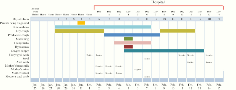
Methods: We report a 55-day-old case with COVID-19 confirmed in China and describe the identification, diagnosis, clinical course, and treatment of the patient, including the disease progression from day 7 to day 11 of illness.

Results: This case highlights that children with COVID-19 can also present with multiple organ damage and rapid disease changes.

Conclusions: When managing such infant patients with COVID-19, frequent and careful clinical monitoring is essential.

Keywords: COVID-19 pneumonia; heart damage; liver injury.


Figures

Figure 1.
Figure 1.
Chest computed tomographic (CT) images of a 55-day-old female infant with COVID-19. NOTE: The scanning condition was low-dose CT with 80-kV tube voltage and 23- to 26-mAs tube current (automatic). (A) The CT images on admission (illness day 6): multiple patchy shadows and ground-glass opacity in the upper and lower lobes of the right lung (black arrow). (B) The CT images on hospital day 4 (illness day 9): compared with day 6 of illness, the lesion progressed and the range widened (gray arrow), accompanied with small consolidation shadow and a few stripe shadows (black arrow). (C) The CT images on on hospital day 11 (illness day 16): compared with day 9 of illness, the inflammation of the right lung was obviously absorbed. Small patchy shadows with increased density and a few strip-like shadows were still observed in the upper lobe of the right lung (black arrow).
Figure 1.
Figure 1.
Chest computed tomographic (CT) images of a 55-day-old female infant with COVID-19. NOTE: The scanning condition was low-dose CT with 80-kV tube voltage and 23- to 26-mAs tube current (automatic). (A) The CT images on admission (illness day 6): multiple patchy shadows and ground-glass opacity in the upper and lower lobes of the right lung (black arrow). (B) The CT images on hospital day 4 (illness day 9): compared with day 6 of illness, the lesion progressed and the range widened (gray arrow), accompanied with small consolidation shadow and a few stripe shadows (black arrow). (C) The CT images on on hospital day 11 (illness day 16): compared with day 9 of illness, the inflammation of the right lung was obviously absorbed. Small patchy shadows with increased density and a few strip-like shadows were still observed in the upper lobe of the right lung (black arrow).
Figure 2.
Figure 2.
Disease course of the infected infant and important information about her parents.

References

    1. Arabi YM, Balkhy HH, Hayden FG, et al. . Middle East respiratory syndrome. N Engl J Med 2017; 376:584–94. - PMC - PubMed
    1. Memish ZA, Al-Tawfiq JA, Assiri A, et al. . Middle East respiratory syndrome coronavirus disease in children. Pediatr Infect Dis J 2014; 33:904–6. - PubMed
    1. Hon KL, Leung CW, Cheng WT, et al. . Clinical presentations and outcome of severe acute respiratory syndrome in children. Lancet 2003; 361:1701–3. - PMC - PubMed
    1. Li AM, Ng PC. Severe acute respiratory syndrome (SARS) in neonates and children. Arch Dis Child Fetal Neonatal Ed 2005; 90:F461–5. - PMC - PubMed
    1. Lu R, Zhao X, Li J, et al. . Genomic characterisation and epidemiology of 2019 novel coronavirus: implications for virus origins and receptor binding. Lancet 2020; 395:1565–74. - PMC - PubMed

Publication types

MeSH terms