

Irisin: Still chasing shadows
- PMID: 32180552
- PMCID: PMC7033458
- DOI: 10.1016/j.molmet.2020.01.016
Irisin: Still chasing shadows
Abstract
Objective: Considerable uncertainty remains regarding the veracity of measuring myokine irisin more than seven years after its original description. Unresolved issues include the nature of transcription of the irisin precursor fibronectin type III domain containing 5 (FNDC5) gene across species, the reliability of irisin levels measured with commercial enzyme-linked immunosorbent assays (ELISAs), and the overall validity of the recently published reference values for human serum measured with quantitative mass spectrometry. We utilized multiple species and measures to evaluate the robustness of commonly used reagents and methods for reporting irisin.
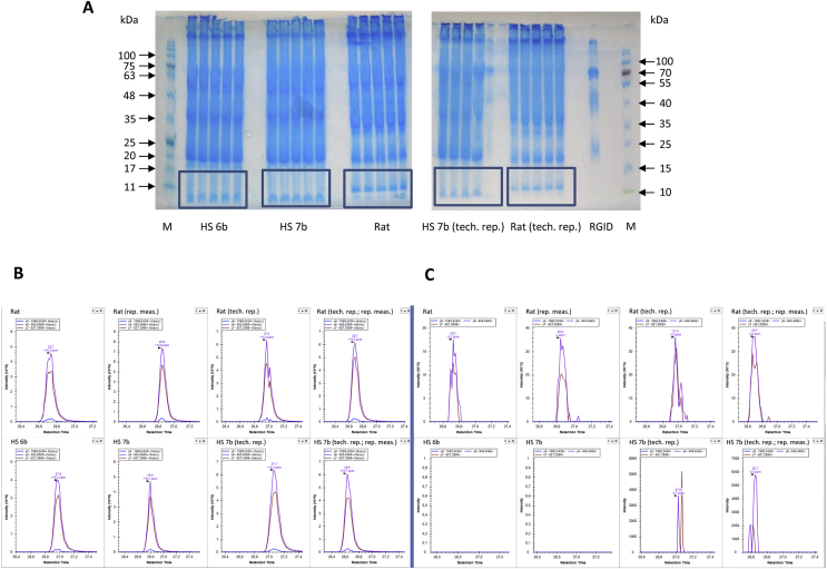
Methods: Amplification of cDNA was used to assess the FNDC5 transcript patterns in humans and mice. The specificity and sensitivity of different irisin antibodies were examined via western blotting. Quantification of circulating native irisin was conducted with mass spectrometry using an absolute quantification peptide for irisin.
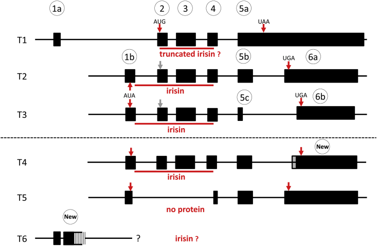
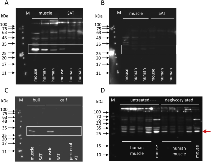
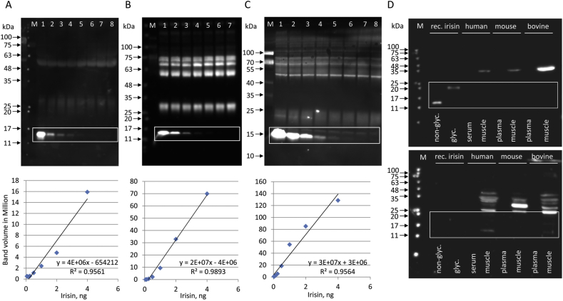
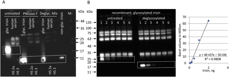
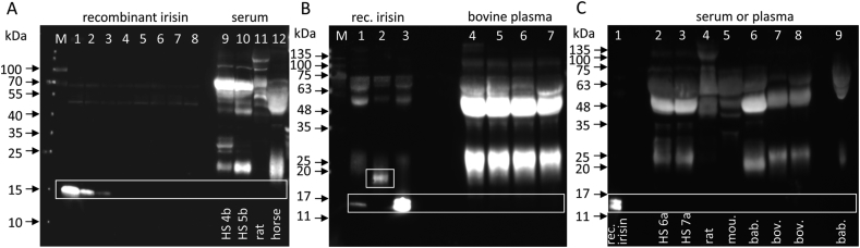
Results: We show that there is a greater transcript diversity of human FNDC5 than currently annotated, but no indication of the expression of transcripts leading to a truncated form of irisin. Available irisin antibodies still bind to patterns of unspecific serum proteins, which compromise reliable measurements of irisin with ELISAs. Absolute quantification of irisin with labeled peptides by mass spectrometry is an advanced method but requires a multi-step sample preparation introducing uncontrollable variations in the measurement.
Conclusion: Our data represent an explicit warning against measuring circulating irisin using available methods. Measuring irisin is akin to chasing shadows.
Keywords: Adipose tissue; FNDC5; Mass spectrometry; Plasma; Skeletal muscle; Transcript.
Copyright © 2020 The Author(s). Published by Elsevier GmbH.. All rights reserved.
Figures
Similar articles
-
FNDC5 expression and circulating irisin levels are modified by diet and hormonal conditions in hypothalamus, adipose tissue and muscle.Sci Rep. 2016 Jul 19;6:29898. doi: 10.1038/srep29898. Sci Rep. 2016. PMID: 27432282 Free PMC article.
-
SMAD3 negatively regulates serum irisin and skeletal muscle FNDC5 and peroxisome proliferator-activated receptor γ coactivator 1-α (PGC-1α) during exercise.J Biol Chem. 2015 Mar 20;290(12):7671-84. doi: 10.1074/jbc.M114.617399. Epub 2015 Feb 3. J Biol Chem. 2015. PMID: 25648888 Free PMC article.
-
Characterization of fibronectin type III domain-containing protein 5 (FNDC5) gene in chickens: Cloning, tissue expression, and regulation of its expression in the muscle by fasting and cold exposure.Gene. 2015 Oct 10;570(2):221-9. doi: 10.1016/j.gene.2015.06.022. Epub 2015 Jun 10. Gene. 2015. PMID: 26072164
-
Is irisin the new player in exercise-induced adaptations or not? A 2017 update.Clin Chem Lab Med. 2018 Mar 28;56(4):525-548. doi: 10.1515/cclm-2017-0674. Clin Chem Lab Med. 2018. PMID: 29127759 Review.
-
An update on the role of irisin in the regulation of endocrine and metabolic functions.Peptides. 2018 Jun;104:15-23. doi: 10.1016/j.peptides.2018.03.018. Epub 2018 Mar 30. Peptides. 2018. PMID: 29608940 Review.
Cited by
-
Effect of Chronic Resistance Training on Circulating Irisin: Systematic Review and Meta-Analysis of Randomized Controlled Trials.Int J Environ Res Public Health. 2021 Mar 3;18(5):2476. doi: 10.3390/ijerph18052476. Int J Environ Res Public Health. 2021. PMID: 33802329 Free PMC article.
-
Cardiometabolic health, visceral fat and circulating irisin levels: results from a real-world weight loss study.J Endocrinol Invest. 2021 Jun;44(6):1243-1252. doi: 10.1007/s40618-020-01415-1. Epub 2020 Sep 6. J Endocrinol Invest. 2021. PMID: 32892317 Free PMC article. Clinical Trial.
-
GLP-1 Induces the Expression of FNDC5 Derivatives That Execute Lipolytic Actions.Front Cell Dev Biol. 2021 Nov 11;9:777026. doi: 10.3389/fcell.2021.777026. eCollection 2021. Front Cell Dev Biol. 2021. PMID: 34869379 Free PMC article.
-
Progress and Challenges in the Biology of FNDC5 and Irisin.Endocr Rev. 2021 Jul 16;42(4):436-456. doi: 10.1210/endrev/bnab003. Endocr Rev. 2021. PMID: 33493316 Free PMC article. Review.
-
Current Evidence of the Role of the Myokine Irisin in Cancer.Cancers (Basel). 2021 May 27;13(11):2628. doi: 10.3390/cancers13112628. Cancers (Basel). 2021. PMID: 34071869 Free PMC article. Review.
References
-
- Timmons J.A., Baar K., Davidsen P.K., Atherton P.J. Is irisin a human exercise gene? Nature. 2012;488:E9–E10. - PubMed
-
- Norheim F., Langleite T.M., Hjorth M., Holen T., Kielland A., Stadheim H.K. The effects of acute and chronic exercise on PGC-1α, irisin and browning of subcutaneous adipose tissue in humans. FEBS Journal. 2014;281:739–749. - PubMed
Publication types
MeSH terms
Substances
LinkOut - more resources
Full Text Sources
Other Literature Sources
