Review
. 2020 Apr:34:27-42.
doi: 10.1016/j.molmet.2019.12.014. Epub 2020 Jan 7.

Mechanisms of insulin resistance related to white, beige, and brown adipocytes

Affiliations
Review

Mechanisms of insulin resistance related to white, beige, and brown adipocytes

Michael P Czech. Mol Metab. 2020 Apr.

Abstract

Background: The diminished glucose lowering effect of insulin in obesity, called "insulin resistance," is associated with glucose intolerance, type 2 diabetes, and other serious maladies. Many publications on this topic have suggested numerous hypotheses on the molecular and cellular disruptions that contribute to the syndrome. However, significant uncertainty remains on the mechanisms of its initiation and long-term maintenance.

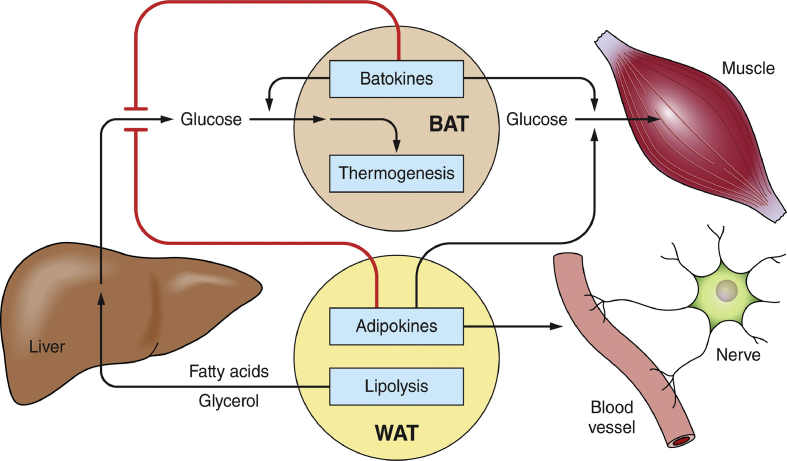
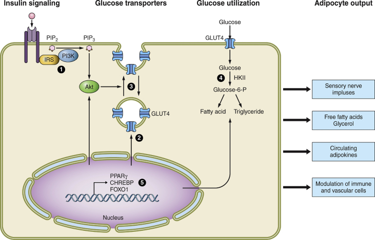
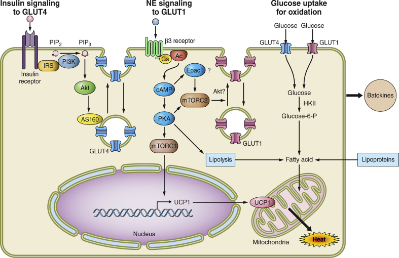
Scope of review: To simplify insulin resistance analysis, this review focuses on the unifying concept that adipose tissue is a central regulator of systemic glucose homeostasis by controlling liver and skeletal muscle metabolism. Key aspects of adipose function related to insulin resistance reviewed are: 1) the modes by which specific adipose tissues control hepatic glucose output and systemic glucose disposal, 2) recently acquired understanding of the underlying mechanisms of these modes of regulation, and 3) the steps in these pathways adversely affected by obesity that cause insulin resistance.

Major conclusions: Adipocyte heterogeneity is required to mediate the multiple pathways that control systemic glucose tolerance. White adipocytes specialize in sequestering triglycerides away from the liver, muscle, and other tissues to limit toxicity. In contrast, brown/beige adipocytes are very active in directly taking up glucose in response to β adrenergic signaling and insulin and enhancing energy expenditure. Nonetheless, white, beige, and brown adipocytes all share the common feature of secreting factors and possibly exosomes that act on distant tissues to control glucose homeostasis. Obesity exerts deleterious effects on each of these adipocyte functions to cause insulin resistance.

Keywords: Adipokines; Adipose tissues; Adrenergic receptors; Bioactive lipids; Glucose tolerance; Lipogenesis; Signaling; Thermogenesis; Uncoupling protein.


Figures

Figure 1
Figure 1
Pathways by which WAT and BAT depots serve as major nodes of systemic metabolic regulation. Adipokines and batokines regulate hepatic lipogenesis and glucose output as well as glucose uptake and disposal by muscle. Secreted factors from adipocytes can also act in a paracrine fashion to regulate other cell types within adipose depots such as vascular cells and nerve fibers. BAT thermogenesis may contribute to systemic glucose disposal and oxidize lipids to lower systemic toxicity. WAT lipolysis in obesity can contribute to fatty acid and glycerol overload in the liver to enhance gluconeogenesis and glucose output. WAT-derived fatty acids also contribute to skeletal muscle insulin resistance (not shown). The combination of the actions of peptides, lipids, small RNA, and other factors from adipocytes plus the released lipolytic products (fatty acids and glycerol) have major influences on local cell types within adipose tissue as well as on distant tissues.
Figure 2
Figure 2
Sites of disrupted functions in white adipocytes due to genetic and diet-induced obesity. At least 5 major insulin-responsive pathways that affect adipocyte metabolism are dysfunctional in obesity and are numbered in the figure. These include 1) insulin signaling elements from insulin receptor to activation of protein kinase Akt, 2) the expression of glucose transporter GLUT4, 3) the membrane trafficking of GLUT4 that enhances its abundance on the plasma membrane in response to insulin, 4) the entry and metabolism of glucose that is converted to fatty acids through de novo lipogenesis and through esterification of glycerol 3-phosphate with fatty acids, and 5) the regulation of expression of many of the proteins and regulators of these pathways through the actions of transcription factors, some of which are themselves responsive to insulin action (for example, CHREBP and FOXO1). These dysfunctions likely all contribute to adipocyte insulin resistance at some stages of acute and chronic obesity, and the degree to which each contributes continue to be actively debated. A key question for current and future studies that is raised by these considerations is the degree to which obesity-altered metabolite levels within adipocytes are able to signal, directly or indirectly, to the outputs at the right of the figure that regulate other cell types within adipose tissue as well as other distant organs such as the brain, liver, muscle, and pancreatic islets.
Figure 3
Figure 3
Mechanisms whereby brown and beige adipocytes take up and utilize glucose to influence whole body insulin sensitivity and glucose tolerance. BAT and beige adipocytes are extremely active in taking up glucose, which may contribute significantly to systemic glucose disposal, at least during cold exposure. Left, insulin signaling to GLUT4: Insulin signaling through Akt to stimulate GLUT4 translocation to the plasma membrane operates as in WAT and BAT, but appears to contribute less to overall glucose uptake in BAT. Obesity does attenuate this pathway in BAT as it does in WAT. Middle, norepinephrine (NE) signaling to GLUT1: Upon stimulation of sympathetic innervation in BAT or WAT, brown and beige adipocytes activate a novel mTORC2-dependent pathway to cause GLUT1 trafficking to the plasma membrane. This significantly stimulates glucose uptake while also activating lipolysis and lipoprotein-based triglyceride hydrolysis to provide fatty acid substrates for uncoupled respiration through UCP1 upregulation. Surprisingly, the mTORC1 complex was found to mediate the expression of UCP1. Right, glucose uptake for oxidation: Glucose uptake in BAT and beige adipocytes is both oxidized and converted to fatty acids by upregulated de novo lipogenesis enzymes during NE stimulation. The newly synthesized fatty acids are thought to be rapidly oxidized in chronic uncoupled respiration. The effects of obesity on GLUT1 translocation and glucose utilization in BAT are less well studied, but the limited data available indicate diminished glucose uptake in BAT in obesity. A key unanswered question is the degree to which altered brown and beige adipocyte metabolites affect the secretion of both beneficial and deleterious batokines that regulate systemic metabolism and the effects of obesity on such regulation.

References

    1. Marks V., Richmond C. 1st ed. Royal Society of Medicine; London: 2007. Insulin murders.
    1. Sweeney J.S. Dietary factors that influence the dextrose tolerance test. A Preliminary Study. Archives of Internal Medicine. 1927;40(6):818–830.
    1. Himsworth H.P. Insulin deficiency and insulin inefficiency. British Medical Journal. 1940;1(4139):719–722. - PMC - PubMed
    1. Yalow R.S., Berson S.A. Immunoassay of endogenous plasma insulin in man. Journal of Clinical Investigation. 1960;39:1157–1175. - PMC - PubMed
    1. Czech M.P. Insulin action and resistance in obesity and type 2 diabetes. Nature Medicine. 2017;23(7):804–814. - PMC - PubMed

Publication types