. 2020 Feb 27:14:30.
doi: 10.3389/fncel.2020.00030. eCollection 2020.

Bi-Directional Communication Between Neurons and Astrocytes Modulates Spinal Motor Circuits

Affiliations

Bi-Directional Communication Between Neurons and Astrocytes Modulates Spinal Motor Circuits

Matthew J Broadhead et al. Front Cell Neurosci. .

Abstract

Evidence suggests that astrocytes are not merely supportive cells in the nervous system but may actively participate in the control of neural circuits underlying cognition and behavior. In this study, we examined the role of astrocytes within the motor circuitry of the mammalian spinal cord. Pharmacogenetic manipulation of astrocytic activity in isolated spinal cord preparations obtained from neonatal mice revealed astrocyte-derived, adenosinergic modulation of the frequency of rhythmic output generated by the locomotor central pattern generator (CPG) network. Live Ca2+ imaging demonstrated increased activity in astrocytes during locomotor-related output and in response to the direct stimulation of spinal neurons. Finally, astrocytes were found to respond to neuronally-derived glutamate in a metabotropic glutamate receptor 5 (mGluR5) dependent manner, which in turn drives astrocytic modulation of the locomotor network. Our work identifies bi-directional signaling mechanisms between neurons and astrocytes underlying modulatory feedback control of motor circuits, which may act to constrain network output within optimal ranges for movement.

Keywords: astrocyte; locomotion; mGlu receptor5; neuromodulation; spinal cord.


Figures

Figure 1
Figure 1
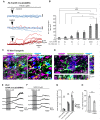
Neuronal activity evokes Ca2+ responses in spinal astrocytes. (A) Schematic of hemisected spinal cord preparations in which simultaneous ventral root recording and Ca2+ imaging of astrocytic activity were performed. (B) Example data from one experiment showing ventral root activity from baseline to the induction of fictive locomotion and Ca2+ imaging traces from four astrocytes (denoted in panel A). Red lines above traces indicate the Ca2+ events detected for analysis. (C) Bar chart showing the duration of astrocytic Ca2+ transients during different phases of the experiment. (D) Bar chart showing the intensity of astrocytic Ca2+ transients during different phases of the experiment. (E) Bar chart showing the frequency of astrocytic Ca2+ transients during different phases of the experiment. (F) Bar chart showing the frequency of astrocytic Ca2+ transients during different phases of the experiment when the hemisected cord was perfused with tetrodotoxin (TTX). (G) Schematic of a spinal cord slice with a simultaneous whole-cell patch-clamp recording from a ventral horn interneuron and Ca2+ imaging of astrocytic activity. (H) Example data from one experiment showing the activation of a spinal cord interneuron (IN), and the Ca2+ transients from three astrocytes (denoted in panel G). (I) Averaged Ca2+ imaging trace from 52 astrocytes from eight separate experiments in response to neuronal stimulation. (J) Bar chart showing the maximum intensity of Ca2+ transients in astrocytes before and after neuronal stimulation. *p < 0.05.
Figure 2
Figure 2
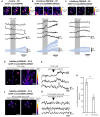
Astrocytic metabotropic glutamate receptor 5 (mGluR5) receptors mediate neuronal-to-astrocyte signaling in the mammalian spinal cord. (A) Example traces from Ca2+ imaging of astrocytes during local applications of vehicle control artificial cerebrospinal fluid (aCSF), N-methyl-D-aspartate (NMDA) and (RS)-2-Chloro-5-hydroxyphenylglycine (CHPG) in the presence of TTX (1 μM). Traces from 10 cells from within the same experiment are shown for each agonist overlayed. Traces are displayed in a red-hue if responsive to the drug, and blue if non-responsive. (B) Bar chart displaying the density of astrocytes that responded to a given pharmacological stimulus. The agonist concentrations and the number of slices (n number) for each agent are listed below the graph. (C) Visualization of mGluR5 receptor expression in glutamine synthetase-labeled astrocytic cell bodies (i), primary leaflet/processes (ii) and finer processes (iii). (D) 3D visualization of mGluR5 localization on astrocytes with orthogonal views of XY, XZ and YZ. Arrows denote cases of colocalization between mGluR5 and glutamine synthetase observed in 3D. (E) Application of NMDA in spinal cord slices from GFAP::Cre;GCAMP6s mice enables the visualization of astrocyte activity in response to neuronal activation. NMDA does not activate astrocytes directly (see Figure 3). (F) Application of NMDA in the presence of mGluR5 antagonist Methyl-6-(phenylethynyl)pyridine hydrochloride (MPEP) shows attenuated responses from astrocytes. (G) Bar chart showing that the degree of astrocytic activation 15 min after NMDA application was significantly reduced when slices were incubated in the mGluR5 antagonist, MPEP. (H) Bar chart showing the number of astrocytes that responded to neuronal activity induced by NMDA was reduced in the presence of MPEP. *p < 0.05, **p < 0.01, ***p < 0.001; ns, not significant.
Figure 3
Figure 3
Pharmacogenetic techniques can manipulate astrocytic Ca2+ activity in the mammalian spinal cord. (A) Ca2+ imaging of GFAP::cre;GCAMP6s astrocytes in response to Clozapine-N-Oxide (CNO). Example (top), and averaged (bottom) traces of Ca2+ imaging from astrocytes during applications of CNO (19 cells for averaged trace). (B) Ca2+ imaging of GFAP::cre;GCAMP6s;hM3Dq astrocytes in response to CNO. Example (top), and averaged (bottom) traces of Ca2+ imaging from astrocytes during applications of CNO (25 cells for averaged trace). (C) Ca2+ imaging of GFAP::cre;GCAMP6s;hM4Di astrocytes in response to CNO. Example (top), and averaged (bottom) traces of Ca2+ imaging from astrocytes during applications of CNO (30 cells for averaged trace). (D) Ca2+ responses in GFAP::cre;GCAMP6s;hM4Di astrocytes in response to glutamate, perfused in TTX to prevent neuronal-mediated responses. (E) Example traces of Ca2+ responses in GFAP::cre;GCAMP6s;hM4Di astrocytes in response to glutamate. (F) Ca2+ responses in GFAP::cre;GCAMP6s;hM4Di astrocytes in response to glutamate, perfused in both TTX and CNO to inhibit astrocyte activity. (G) Example traces of Ca2+ responses in GFAP::cre;GCAMP6s;hM4Di astrocytes showing no response to glutamate due to pharmacogenetic inhibition with CNO. (H) Bar chart displaying the number of astrocytes responding to glutamate in GFAP::cre;GCAMP6s;hM4Di spinal cord slices under control conditions and when perfused with CNO to inhibit astrocytes. **p < 0.01.
Figure 4
Figure 4
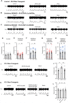
Astrocytes exert purinergic modulation of locomotion in response to mGluR5 dependent activation. (A) Example raw (top) and rectified/integrated (bottom) traces of pharmacologically induced fictive locomotor bursts recorded from in vitro spinal cord preparations from control non-transgenic neonatal mice. The burst frequency, duration and amplitude were analyzed in control conditions, in the presence of CNO and following the washout of CNO. (B) Pharmacologically induced fictive locomotor bursts recorded from a GFAP::cre;hM3Dq (excitatory DREADD) neonatal mouse spinal cord in vitro during control conditions, in the presence of CNO and following the washout of CNO. (C) Pharmacologically induced fictive locomotor bursts recorded from a GFAP::cre;hM4Di (inhibitory DREADD) neonatal mouse spinal cord in vitro during control conditions, in the presence of CNO and following the washout of CNO. (D) Bar chart displaying the frequency of fictive locomotor bursts in control, non-transgenic mouse spinal cords in response to CNO. (E) Bar chart displaying the frequency of fictive locomotor bursts in excitatory DREADD mouse spinal cords in response to CNO. (F) Bar chart displaying the frequency of fictive locomotor bursts in inhibitory DREADD mouse spinal cords in response to CNO. (G) Bar chart displaying the frequency of fictive locomotor bursts in excitatory DREADD mouse spinal cords in response to CNO when perfused with 8-Cyclopentyl-1,3-dipropylxanthine (DPCPX) throughout to block the adenosine A1 receptor. (H) Bar chart displaying the frequency of fictive locomotor bursts in inhibitory DREADD mouse spinal cords in response to CNO when perfused with DPCPX throughout to block the adenosine A1 receptor. (I) Example traces of fictive locomotion recorded from in vitro spinal cord preparations from control non-transgenic neonatal mouse spinal cords, showing the effects of a 20 min application of the A1 receptor antagonist, DPCPX. (J) Bar chart showing that the frequency of fictive locomotion is significantly increased with the application of DPCPX. (K) Example traces of fictive locomotion recorded from in vitro spinal cord preparations, showing blockade of the effects of the application of DPCPX when in the presence of MPEP. (L) Bar chart showing that DPCPX has no effect on the frequency of fictive locomotion when spinal cords are also incubated in MPEP. *p < 0.05, ns; not significant.

References

    1. Acevedo J., Santana-Almansa A., Matos-Vergara N., Marrero-Cordero L. R., Cabezas-Bou E., Díaz-Ríos M. (2016). Caffeine stimulates locomotor activity in the mammalian spinal cord via adenosine A1 receptor-dopamine D1 receptor interaction and PKA-dependent mechanisms. Neuropharmacology 101, 490–505. 10.1016/j.neuropharm.2015.10.020 - DOI - PMC - PubMed
    1. Acton D., Broadhead M. J., Miles G. B. (2018). Modulation of spinal motor networks by astrocyte-derived adenosine is dependent on D1-like dopamine receptor signaling. J. Neurophysiol. 120, 998–1009. 10.1152/jn.00783.2017 - DOI - PMC - PubMed
    1. Acton D., Miles G. B. (2015). Stimulation of glia reveals modulation of mammalian spinal motor networks by adenosine. PLoS One 10:e0134488. 10.1371/journal.pone.0134488 - DOI - PMC - PubMed
    1. Acton D., Miles G. B. (2017). Gliotransmission and adenosinergic modulation: insights from mammalian spinal motor networks. J. Neurophysiol. 118, 3311–3327. 10.1152/jn.00230.2017 - DOI - PMC - PubMed
    1. Allen N. J. (2019). Star power: astrocytes regulate behavior. Cell 177, 1091–1093. 10.1016/j.cell.2019.04.042 - DOI - PubMed

LinkOut - more resources