

Right-sided obstructed hemivagina ipsilateral renal agenesis (OHVIRA): A case report
- PMID: 32181147
- PMCID: PMC7063130
- DOI: 10.1016/j.crwh.2020.e00185
Right-sided obstructed hemivagina ipsilateral renal agenesis (OHVIRA): A case report
Abstract
Introduction: Obstructed hemivagina ipsilateral renal agenesis (OHVIRA) is a rare anomaly of the urogenital system. The characteristic triad of this syndrome, which was initially reported in 1950, is didelphys uterus, obstructed hemivagina, and ipsilateral renal agenesis (Embrey, 1950 [1]).
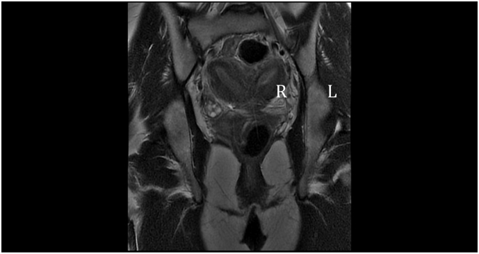
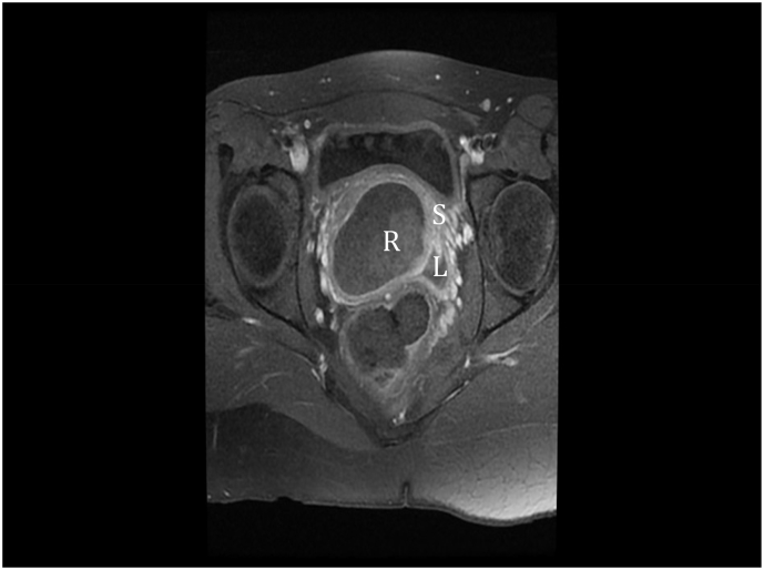
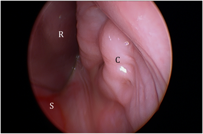
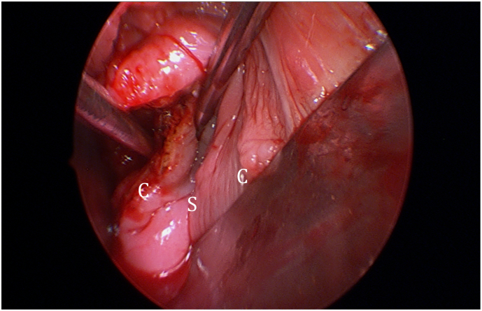
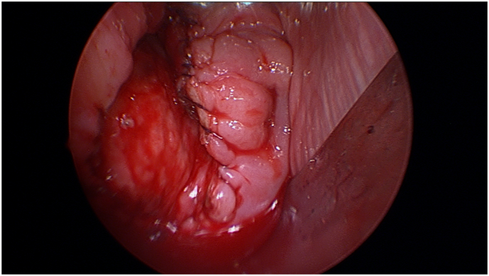
Case: A 17-year-old girl was referred with a 6-month history of offensive vaginal discharge. Magnetic resonance imaging (MRI) established the diagnosis of OHVIRA. She underwent surgery for drainage of the hematocolpos and excision of the vaginal septum followed by an uncomplicated recovery and the patient had normal menstrual cycles after surgery.
Conclusion: There should be a high suspicion of OHVIRA syndrome when encountering adolescent patients with non-specific abdominal or pelvic symptoms.
Keywords: Didelphys and bicollis; MRI, magnetic resonance imaging; Mullerian anomaly; OHVIRA; OHVIRA, Obstructed hemivagina ipsilateral renal agenesis; Obstructed hemivagina; Renal agenesis; USS, ultrasound scan; Urogenital malformation.
© 2020 The Authors.
Figures
References
-
- Shavell V.I., Montgomery S.E., Johnson S.C., Diamond M.P., Berman J.M. Complete septate uterus, obstructed hemivagina, and ipsilateral renal anomaly: pregnancy course complicated by a rare urogenital anomaly. Arch. Gynecol. Obstet. 2009;280(3):449–452. - PubMed
-
- Candiani G.B., Fedele L., Candiani M. Double uterus, blind hemivagina, and ipsilateral renal agenesis: 36 cases and long-term follow-up. Obstet. Gynecol. 1997;90(1):26–32. - PubMed
-
- Smith N.A., Laufer M.R. Obstructed hemivagina and ipsilateral renal anomaly (OHVIRA) syndrome: management and follow-up. Fertil. Steril. 2007;87(4):918–922. - PubMed
Publication types
LinkOut - more resources
Full Text Sources
