Review
. 2020 Feb 28:8:112.
doi: 10.3389/fchem.2020.00112. eCollection 2020.

Probing of Nucleic Acid Structures, Dynamics, and Interactions With Environment-Sensitive Fluorescent Labels

Affiliations
Review

Probing of Nucleic Acid Structures, Dynamics, and Interactions With Environment-Sensitive Fluorescent Labels

Benoît Y Michel et al. Front Chem. .

Abstract

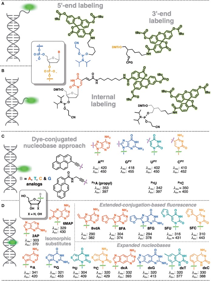
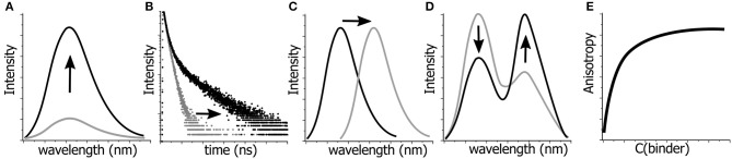
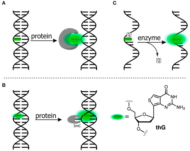
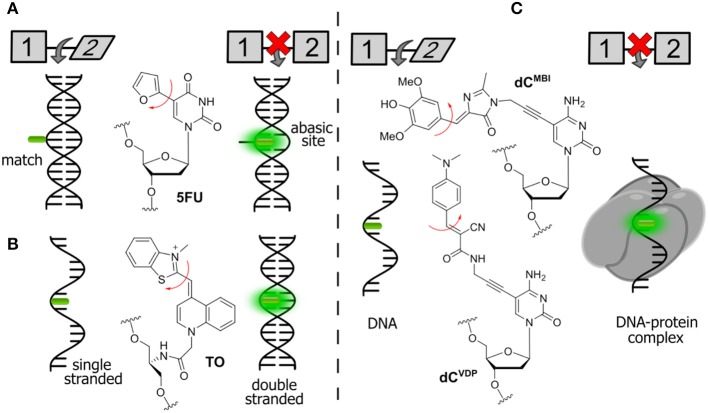
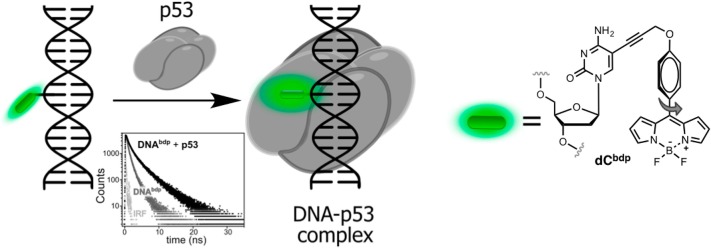
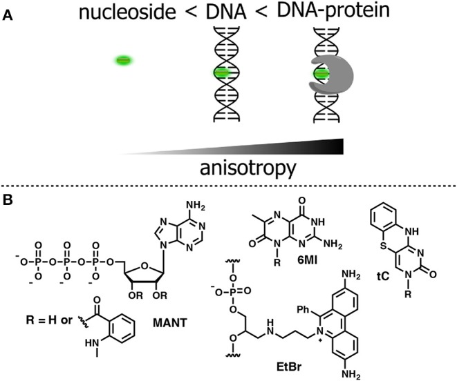
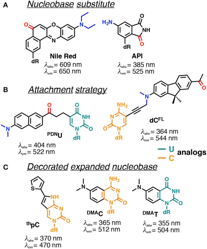
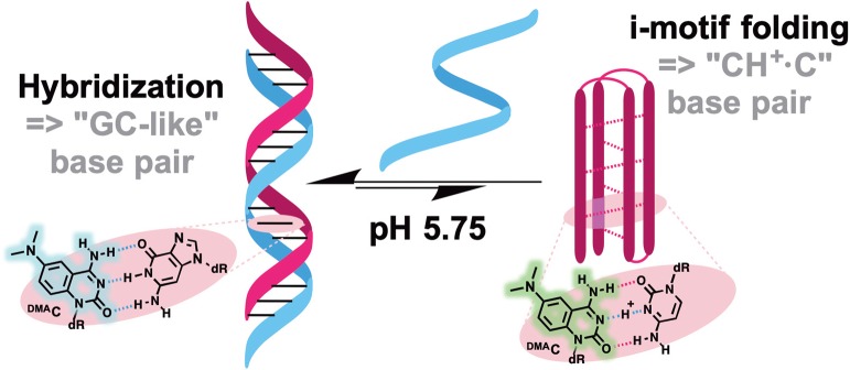
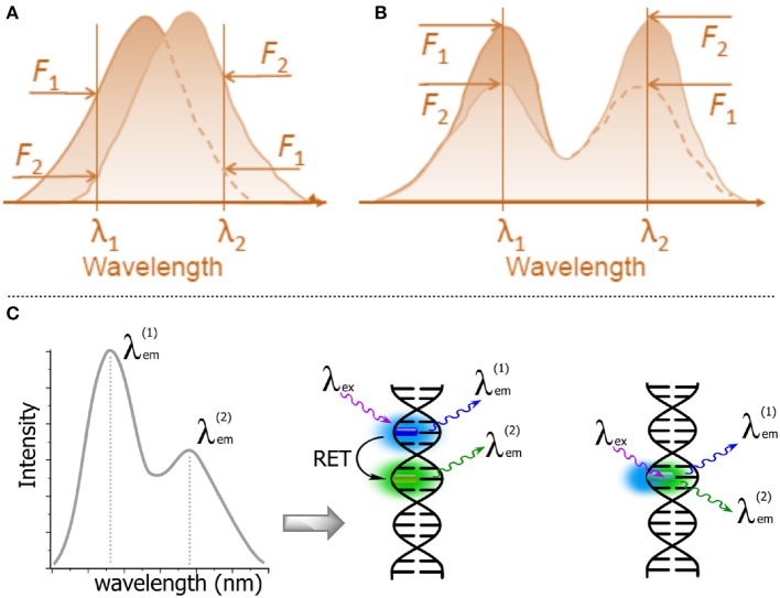
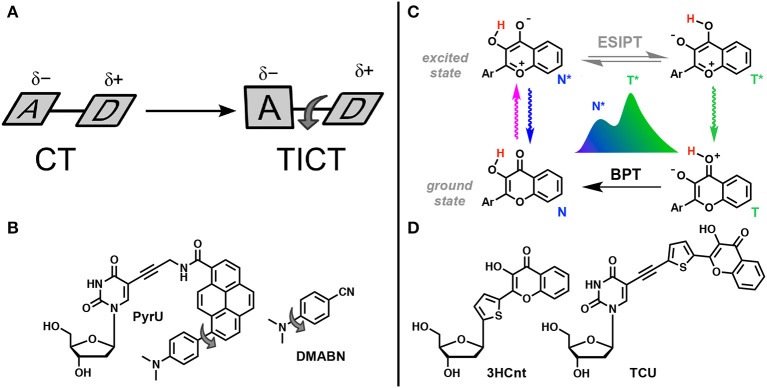
Fluorescence labeling and probing are fundamental techniques for nucleic acid analysis and quantification. However, new fluorescent probes and approaches are urgently needed in order to accurately determine structural and conformational dynamics of DNA and RNA at the level of single nucleobases/base pairs, and to probe the interactions between nucleic acids with proteins. This review describes the means by which to achieve these goals using nucleobase replacement or modification with advanced fluorescent dyes that respond by the changing of their fluorescence parameters to their local environment (altered polarity, hydration, flipping dynamics, and formation/breaking of hydrogen bonds).

Keywords: emissive nucleobase; fluorescence sensing; nucleoside analog; probing interactions; probing nucleic acids.


Figures

Figure 1
Figure 1
Main strategies for covalent fluorescence labeling of NAs: via a flexible tether either at the 3′/5′-end (A), or at an internal position (B) illustrated here by amidite building blocks bearing a fluorescein (Teo and Kool, ; Lavis and Raines, 2014); (C) through a short and rather rigid linker for extra-helical probing depicted by pyrene, an aromatic polycyclic dye connected to the pyrimidine C5 and purine C8 positions (Rist et al., ; Okamoto et al., , ; Saito et al., ; Hwang et al., ; Seo et al., ; Seo and Kim, ; Østergaard and Hrdlicka, 2011) and as a nucleobase surrogate for intra-helical probing (D) exemplified by selected isomorphic base mimics (Hawkins, ; McCoy et al., ; Park et al., ; Jones and Neely, 2015) and nucleobases made emissive by ring expansion (Gao et al., ; Liu et al., ; Krueger et al., ; Srivatsan et al., ,; Shin et al., 2011) or extension of conjugation (Gaied, ; Greco and Tor, , ; Srivatsan and Tor, ,; Sinkeldam et al., ; Greco et al., 2009). Excitation and emission wavelengths were given in nm.
Figure 2
Figure 2
Possibilities for monitoring the changes in molecular structures, dynamics and interactions by fluorescence spectroscopy. Variations in: (A) fluorescence intensity, (B) fluorescence lifetimes, (C) position of the emission maximum, i.e., emission color, (D) ratio of intensities at two wavelengths, and (E) fluorescence anisotropy can be detected and quantified.
Figure 3
Figure 3
Single-dye hybridization probes for the detection of SNPs. (A) Pyrrolocytosine nucleobase allowing mismatch discrimination by a fluorescent turn-on response; and (B) quencher-free molecular beacon approach revealing match cases by a light-up signal.
Figure 4
Figure 4
Fluorogenic probes for sensing interactions with enzymes based on NAs labeled with a single FNA: (A) ssDNA probe for endo- and exonucleases; (B) ds-RNA/DNA probe for RNase H; and (C) hairpin RNA probe for adenosine deaminase.
Figure 5
Figure 5
FNAs as a base mimic and stacking-sensitive reporter (A,B) for sensing single-base flipping; (C) for sensing DNA damage and repair. Q, quencher being either U or 1-methyladenine.
Figure 6
Figure 6
Selected examples of FNAs featuring segmental mobility and their applications in sensing. (A) Furan-decorated uridine used as an abasic site sensor; (B) peptide-type FNA becoming emissive by forced intercalation; and (C) bearing flexible dyes, cytidine FNAs affording fluorescence enhancement upon protein interactions.
Figure 7
Figure 7
Sequence-specific binding of the transcription factor p53 to DNA sensed via lifetime experiments. Fluorescence decays of labeled DNA before (light gray) and after (dark gray) binding to p53.
Figure 8
Figure 8
(A) Relative changes in anisotropy for increasing molecular sizes of the dye-containing complex. (B) FNA suitable for anisotropy-based sensing.
Figure 9
Figure 9
Selected examples of solvatofluorochromic FNAs demonstrating intramolecular charge transfer (ICT): (A) employed as nucleobase surrogates; (B) covalently and flexibly attached to a pyrimidine; and (C) mimicking nucleobases by an appropriate expansion. dR indicates 2'-deoxyribosyl; donor and acceptor groups are depicted in blue and red, respectively (Barawkar and Ganesh, ; Okamoto et al., ; Tainaka et al., ; Noé et al., ; Riedl et al., ; Weinberger et al., ; Mata and Luedtke, ; Mata et al., 2016). Excitation and emission wavelengths were given in nm.
Figure 10
Figure 10
pH-induced i-motif folding monitored by color change of DMAC fluorescence.
Figure 11
Figure 11
Schematic representations of λ-ratiometric sensing recorded at two fixed wavelengths for a single fluorescent probe. (A) The spectral shift defines the sensing response (e.g., Nile Red). (B) The decrease of intensity of one band λ1 and the concomitant increase of intensity at a different band λ2 define the sensing response. Calculating the change in the intensity ratio F1)/F2) enables quantitative measurements (Demchenko, 2013). (C) Dual emission: two-dyes (e.g., exhibiting resonance energy transfer, RET) vs. a one-dye approach based on the generation of a new band for the recording of a ratiometric signal.
Figure 12
Figure 12
TICT vs. ESIPT. (A) Schematic representation of geometrical arrangements of normal charge transfer (CT) and twisted intramolecular charge transfer (TICT) excited states (Rettig, 1986). (B) FNA containing fluorophore undergoing TICT reported by Okamoto et al. (2005b). (C) Tautomeric equilibrium explaining the dual-band emission of 3-hydroxychromones. ESIPT, excited-state intramolecular proton transfer; BPT, back proton transfer; N* and T* states for normal and tautomer excited forms, respectively. (D) Dual-emissive nucleoside analogs based on 3-hydroxychromones (Dziuba et al., ; Barthes et al., 2015).

References

    1. Allan B. W., Reich N. O. (1996). Targeted base stacking disruption by the EcoRI DNA methyltransferase. Biochemistry 35, 14757–14762. 10.1021/bi9615708 - DOI - PubMed
    1. Asseline U. (2006). Development and applications of fluorescent oligonucleotides. Curr. Org. Chem. 10, 491–518. 10.2174/138527206776055349 - DOI
    1. Bahr M., Valis L., Wagenknecht H.-A., Weinhold E. (2007). DNA labelling topologies for monitoring DNA-protein complex formation by fluorescence anisotropy. Nucleos. Nucleot. Nucl. 26, 1581–1584. 10.1080/15257770701547347 - DOI - PubMed
    1. Barawkar D. A., Ganesh K. N. (1995). Fluorescent d(CGCGAATTCGCG): characterization of major groove polarity and study of minor groove interactions through a major groove semantophore conjugate. Nucl. Acids Res. 23, 159–164. 10.1093/nar/23.1.159 - DOI - PMC - PubMed
    1. Barlev A., Sen D. (2013). Catalytic DNAs that harness violet light to repair thymine dimers in a DNA substrate. J. Am. Chem. Soc. 135, 2596–2603. 10.1021/ja309638j - DOI - PubMed

LinkOut - more resources