

Characterization of Disease Progression in the Initial Stages of Retinopathy in Type 2 Diabetes: A 2-Year Longitudinal Study
- PMID: 32181799
- PMCID: PMC7401457
- DOI: 10.1167/iovs.61.3.20
Characterization of Disease Progression in the Initial Stages of Retinopathy in Type 2 Diabetes: A 2-Year Longitudinal Study
Abstract
Purpose: To characterize 2-year changes occurring in neurodegeneration, edema, and capillary dropout in nonproliferative diabetic retinopathy.
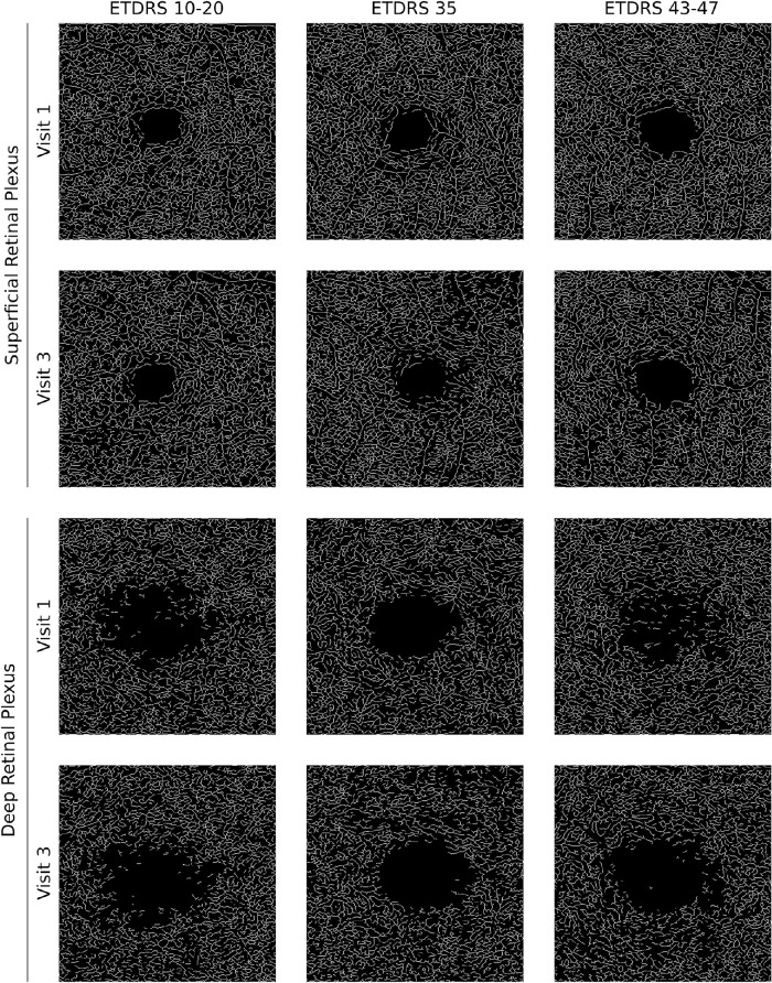
Methods: Two-year prospective longitudinal observational cohort of eyes/patients with type 2 diabetes using spectral domain optical coherence tomography (SD-OCT) and optical coherence tomography angiography (OCTA). Eyes were examined three times with intervals of 1 year. Thickness of the full retina and layer-by-layer measurements were used to identify edema or neurodegeneration. OCTA vessel density maps of the retina were used to identify capillary dropout. Early Treatment Diabetic Retinopathy Study (ETDRS) classification was performed using the seven-field ETDRS protocol.
Results: A total of 62 eyes from 62 patients with diabetes were followed for 2 years. After verification for image quality, a total of 44 eyes from 44 patients (30% women) aged 52 to 80 years were retained for data analysis. There were 18 eyes with ETDRS grades 10 to 20, 17 eyes with ETDRS grade 35, and 9 eyes with ETDRS grades 43 to 47. During the 2-year follow-up period, there was a progressive increase in capillary dropout, whereas edema and neurodegeneration remained stable. In multivariate analysis, considering a model adjusted for age, sex, hemoglobin A1C, visual acuity, and diabetes duration, vessel density remained significantly different between Diabetic Retinopathy Severity Scale groups (Wilks' λ = 0.707; P = 0.015) showing association with disease progression.
Conclusions: Capillary dropout increased in a period of 2 years in eyes with minimal, mild, and moderate diabetic retinopathy, whereas the presence of edema and neurodegeneration remained stable.
Conflict of interest statement
Disclosure:
Figures

References
-
- Wang RK, Jacques SL, Ma Z, Hurst S, Hanson SR, Gruber A. Three dimensional optical angiography. Opt Express. 2007; 15: 4083. - PubMed
-
- Soares M, Neves C, Marques IP, et al.. Comparison of diabetic retinopathy classification using fluorescein angiography and optical coherence tomography angiography. Br J Ophthalmol. 2017; 101: 62–68. - PubMed
-
- Bandello F, Tejerina AN, Vujosevic S, et al.. Retinal layer location of increased retinal thickness in eyes with subclinical and clinical macular edema in diabetes type 2. Ophthalmic Res. 2015; 54: 112–117. - PubMed
Publication types
MeSH terms
LinkOut - more resources
Full Text Sources
Medical
